Etiology
Transitional Cell Cancer
In the United States, bladder cancer is reportedly the fourth most common malignancy. The vast majority of bladder neoplasms arise from the epithelium, with urothelial (transitional cell) carcinoma accounting for 90% of cases. Squamous cell carcinoma is rare and accounts for 2% to 15% of bladder cancers. The least common is adenocarcinoma, which may be primary or metastatic to the bladder.
Fibroma
Fibroma (leiomyoma) is the most common benign tumor of the bladder, although it represents only 0.4% of all bladder neoplasms. It is most prevalent among women in the third to fifth decades of life.
Endometriosis
Endometriosis occurs as a fusiform mass in the bladder wall and is covered in the section on endometriosis.
Diffuse Bladder Wall Thickening
Diffuse bladder wall thickening is seen in cases of severe chronic cystitis or in chronic bladder outlet obstruction where the bladder becomes trabeculated (more common in males with large prostates).
Findings Specifically Related to the Ureteral Orifice
- •
Ureterocele
- •
Ureteral reimplantation site
- •
Stone at the ureteropelvic junction (UPJ) with edema of ureteral orifice
Urethral Diverticula
Urethral diverticula occur just under the bladder along the urethra. They can be quite painful, especially when the patient voids.
Other Bladder Masses
There are many benign tumors such as paraganglioma, plasmacytoma, hemangioma, neurofibroma, and lipoma that can occasionally (rarely) occur in the bladder. Malignant neoplasms reported in the bladder include rhabdomyosarcoma, leiomyosarcoma, lymphoma, osteosarcoma, and metastatic tumors such as melanoma.
Ultrasound Findings
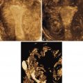
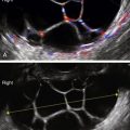
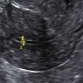
Bladder masses are typically located in the bladder wall. Because these arise from the submucosal portion of the bladder wall, they typically appear as smooth intramural lesions. Transitional cell carcinoma is a focal mucosal lesion, which is fungating and extends into the lumen of the bladder with an irregular surface. Color Doppler usually reveals abundant blood flow, as with other pelvic malignancies. Bladder wall lesions are typically fusiform with an intact mucosal surface and focally widen the wall of the bladder. Endometriosis of the bladder wall (likely the most common diagnosis in gynecologic patients) has spotty blood flow by Doppler and a smooth inner and outer wall. Lesions involving the ureteral orifice can be cystic, such as ureteroceles (which are usually asymptomatic); in the case of reimplantations, there will be a surgical history.
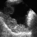
Urethral diverticula are complex masses along the urethra, just under the bladder (see Vaginal Masses ). These may indent the floor of the bladder and be quite painful during the ultrasound. They are best seen by placing the transducer on the perineum and looking cephalad toward the bladder. They are usually cystic with varying amounts of solid area and calcification, depending on chronicity of the lesion.
Related posts:


Stay updated, free articles. Join our Telegram channel

Full access? Get Clinical Tree


