and Lori Ann Lewis1
(1)
Clinical Research Center of North Texas, Denton, TX, USA
Abstract
Most full-size central DXA densitometers have software options to determine body composition from a total body bone density study. This application was first developed for dual photon absorptiometers, but the almost 1-h scan time made such measurements clinically impractical. In contrast, today’s DXA devices can perform total body scans in a matter of minutes. In spite of this dramatic improvement in speed, body composition assessment with DXA remains an underutilized application.
Most full-size central DXA densitometers have software options to determine body composition from a total body bone density study. This application was first developed for dual photon absorptiometers, but the almost 1-h scan time made such measurements clinically impractical. In contrast, today’s DXA devices can perform total body scans in a matter of minutes. In spite of this dramatic improvement in speed, body composition assessment with DXA remains an underutilized application.
The assessment of body composition is much different from the measurement of weight, although certainly the two are related. The assessment of body composition is concerned with the percentage and distribution of fat and lean tissue in the body. Although many studies have associated extremes of weight with disease states, it is increasingly recognized that the percentage and distribution of fat and lean tissue are as or even more important than total body weight in various disease states. A brief review of other techniques used to assess body composition is useful in order to appreciate the unique aspects of body composition analysis with DXA.
The Body Mass Index
The body mass index (BMI) is only a first step in the assessment of body composition, but it goes beyond the simple measurement of total body weight. The BMI relates the patient’s weight to their height, using the following formula:


(14.1)
For example, if a woman weighs 120 lbs and has a height of 62 in., her BMI is


(14.2)

(14.3)
When using the metric system to measure height and weight, the formula changes to


(14.4)
In the example given above, the patient’s weight of 120 lbs and height of 62 in. converts1 to 54.53 kg and 1.575 m. The formula for BMI thus becomes


(14.5)

(14.6)
The very slight differences in results are due to the effects of converting the original values to the metric system and rounding. However, in both cases, the BMI is essentially 22. The formula for calculating BMI is also called Quetelet’s formula or index [1].
In 1995, the World Health Organization (WHO) used the BMI to define obesity in adults [2]. In 1998, the WHO also defined categories of risk based primarily on premature mortality for any given BMI [3]. These divisions are shown in Table 14-1. The criteria apply to both women and men. Some DXA body composition software will calculate and plot the patient’s BMI on a graphic scale that indicates the WHO classification. An example is shown in Fig. 14-1.
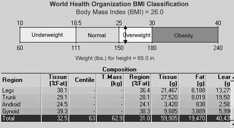
Table 14-1
The World Health Organization criteria for obesity and risk for premature mortality based on the BMI for adults aged 20 and older
Category | BMI (kg/m2) | Risk |
|---|---|---|
Underweight | <18.50 | Low (but other comorbidities increased) |
Normal | 18.50–24.99 | Average |
Overweight | ≥25.00 | |
Preobese | 25.00–29.99 | Increased |
Obese class I | 30.00–34.99 | Moderate |
Obese class II | 35.00–39.99 | Severe |
Obese class III | ≥40.00 | Very severe |

Fig. 14-1.
Ancillary body composition data from a GE Lunar Prodigy body composition study in which the patient’s BMI has been plotted on a scale representing the World Health Organization classification system for obesity.
Although the BMI is an improvement over the assessment of weight alone, it does not address the actual percentage of total body fat or the distribution of fat in the body. The BMI obviously cannot distinguish the contribution to weight from either lean or fat tissue. As a consequence, individuals with a large muscle mass may actually have a BMI that results in a classification of overweight or obese, when clearly they are not. More sophisticated methods for body composition are required to quantify the percentages of fat and lean tissue and the distribution of each within the body.
Body Composition Methods
The body can be divided into two major compartments: fat and fat-free. The fat-free compartment can be further divided into water, protein, and mineral. The various techniques used to measure body composition are characterized by the number of compartments that they measure. A 2-compartment method measures fat and fat-free mass. If a method is described as a 3- or 4-compartment method, it is measuring fat mass and 2 or 3 of the components of the fat-free mass, respectively.
The traditional “gold standard” for measuring body composition has been a 2-compartment technique called underwater weighing (UWW).2 Other 2 compartment methods include the measurement of skinfold thickness, bioelectrical impedance, and air displacement plethysmography. Near-infrared interactance (NIR) and DXA body composition assessments are 3-compartment methods.
2-Compartment Body Composition Measurement Techniques
Underwater Weighing
Underwater weighing (UWW) is one of the most widely used methods for assessing body composition and, as noted earlier, has long been considered the gold standard for these types of techniques. The principle behind UWW is that there is an inverse relationship between body fat and body density. That is, as one goes up, the other goes down. The major assumption in UWW is that the densities of the fat and fat-free mass in the body are constant [4]. It is generally agreed that fat mass has a density of 0.9 g/cm3, but the density of the fat-free mass is controversial. In the past, the density of the fat-free mass was assumed to be a relatively constant 1.1 g/cm3. This does not appear to be true. Recall that the fat-free mass consists of water, protein, and bone mineral. Variations in any of these components may cause the density of the fat-free mass to change. Although the bone mineral is a small percentage of the fat-free mass, it contributes significantly to the density of the fat-free mass. As a consequence, changes in the amount of bone mineral have a greater influence on the total density of the fat-free mass than might otherwise be expected. This creates potential inaccuracies in the measurement of total body fat with UWW in which the amount of bone mineral is assumed, rather than measured. If the density of the fat-free mass is lower because of a decreased total body bone mineral content, the total body percent fat will be overestimated by UWW [5]. The converse is also true; if the density of the fat-free mass is higher because the total body bone mineral is greater than assumed, UWW will underestimate total body percent fat.
The technique calls for the complete submersion of the individual in a tank of water. The individual is asked to exhale as much as possible, and then, while holding their breath, they are submerged and weighed underwater. Under ideal circumstances, no clothing of any kind is allowed for the measurement. This requirement is often waived, but this will affect the accuracy of the measurement to a small extent as well. The amount of air left in the lungs after completely exhaling, the residual lung volume, must be measured since this is expected to provide some buoyancy to the body while underwater. In some institutions, the residual lung volume is estimated, rather than measured, using equations that are specific for age, height, and gender.
The person’s weight underwater can be calculated based on the Archimedes principle that there is a buoyant counterforce equal to the weight of the water that is displaced by the body. Consequently, the weight of the body underwater reflects the weight of the body minus the weight of the fluid that is displaced by the volume of the body. When combined with knowledge of the residual lung volume, the volume and density of the body can be estimated. The classic equation used to calculate the density of the fat mass from the total body density is the equation from Siri [6] in which
![$$ F=\left[4.95\times \left(\frac{1}{D}\right)-4.50\right],$$](/wp-content/uploads/2016/04/A145815_3_En_14_Chapter_Equ00147.gif)
where F is the density of the fat mass and D is the density of the total body. In recent years, however, equations have been developed for the calculation of fat density that is age and gender specific.
![$$ F=\left[4.95\times \left(\frac{1}{D}\right)-4.50\right],$$](/wp-content/uploads/2016/04/A145815_3_En_14_Chapter_Equ00147.gif)
(14.7)
Skinfold Measurements
Body fat can be estimated from multiple measurements of skinfold thickness. This is also considered a two-compartment method. In this method, it is assumed that the distribution of subcutaneous fat and internal fat is similar in everyone. This is a major assumption that is not necessarily valid. Nevertheless, there are equations that allow the calculation of percent body fat from skinfold thickness for men and women.
The technique requires that the skinfold thickness be measured at multiple sites. There are calipers made specifically for this purpose such as the Lange Skinfold Caliper and the Harpenden Skinfold Caliper. The number of sites measured varies from 3 to 7. The 7-site method calls for measurements of skinfold thickness at the chest, triceps, subscapular region, axilla, suprailiac region, abdomen, and thigh. The more commonly used 3-site method calls for measurements at the chest, abdomen, and thigh in men and at the triceps, thigh, and suprailiac region in women.
The measurement of skinfold thickness at the various sites is then used in an equation to calculate the total body density. Equations have been developed by Durnin and Womersley [7] as well as Jackson and Pollock [8, 9] for this purpose, although Jackson and Pollock also combined gluteal or waist circumference with skinfold measurements to calculate body density. The classic formula to calculate total body density from the 7-skinfold thickness measurement in men is

where D is the total body density, X is the sum of the 7 skinfolds in mm, and A is the age in years. In women, the corresponding formula is


(14.8)

(14.9)
For the 3-skinfold thickness measurement, specific equations also exist for men and women. For men

where D is again the total body density, X is now the sum of the chest, abdomen, and thigh skinfolds, and A is age in years. For women, the corresponding equation is

where X is the sum of the triceps, thigh, and suprailiac skinfolds. Variations of these equations from other authors exist as well. With any of these equations, once the total body density is known, an equation such as (14.7) in this chapter from Siri [6] can be used to calculate the percentage of total body fat. Today, these calculations are readily performed by computer programs. The accuracy and reproducibility of skinfold measurements are highly dependent on the skill of the individual making the measurements.

(14.10)

(14.11)
Bioelectrical Impedance Analysis
Bioelectrical impedance analysis (BIA) is also considered a 2-compartment method. In this technique, the individual commonly stands barefoot on a metal foot plate from which an extremely low-voltage electric current is sent up one leg and then down the other. The individual may also be asked to lie on a table with electrodes connected to both legs after which an undetectable low-voltage electric current is transmitted through the body from the electrodes. Because fat is a very poor conductor of electricity and water, a component of the fat-free mass, is a very good conductor of electricity, the resistance to the electric current can be used to estimate the percent body fat.
BIA is a popular method, often found in health clubs because of its ease of use and short measurement time (less than 1 min). It is generally considered to overestimate body fat in lean individuals and underestimate body fat in obese individuals. Because BIA results are highly dependent on total body water, the hydration status of the individual can profoundly affect the results. As a consequence, individuals are advised to abstain from eating or drinking 4 h prior to the test, avoid exercise within 12 h of the test, and abstain from alcohol for 48 h prior to test. Immediately prior to testing, the individual is asked to empty their bladder. Diuretic use can also affect the results obtained with BIA.
Air Displacement Plethysmography
Air displacement plethysmography can be considered analogous to underwater weighing, except that it is the displacement of air instead of the displacement of water that is used to measure the density and volume of the body. One technique using air displacement is called the BOD POD® (COSMED USA, Inc., Chicago, IL). This is an enclosed, egg-shaped, capsule-like structure. An individual undergoing the measurement sits very still within the capsule and breathes quietly during the 5–8 min measurement. A swim cap and tight fitting clothing are generally worn. As in underwater weighing, it is necessary to determine the residual lung volume, by either measurement or estimation. The equipment is relatively expensive which limits its availability, although less expensive models have become available. Measurements of body fat with the BOD POD® have been reported to be highly correlated with those made by underwater weighing [10]. In a review of the published literature from December 1995 to August 2001, the average percent body fat in children and adults differed by less than 1 % between the BOD POD® and UWW and by less than 1 % between the BOD POD® and DXA in adults and less than 2 % in children [11].
3-Compartment Body Composition Measurement Techniques
Near-Infrared Interactance
Near-infrared interactance (NIR) for body composition analysis relies on the principles of infrared spectroscopy. A computerized spectrophotometer is used in this method. A probe which emits an infrared light is placed on the body. The infrared light passes through the fat and muscle and is then reflected back to the probe. The reflected light is quantified and used to calculate body density. The method is considered quite safe and is fast, convenient, and inexpensive. It requires no particular patient preparation. The NIR unit itself is generally small and portable. The Futrex®-6100 and Futrex®-5500 (Futrex Inc., Gaithersburg, MD) are commercial brands of NIR body composition devices. The accuracy of NIR has been compared to skinfolds and BIA using UWW as the reference technique [12–14]. In general, the results obtained from skinfold measurements, BIA, and NIR were all highly correlated with the results from UWW. At the extremes of weight, skinfold measurements appeared to be more accurate than BIA or NIR when UWW was used as the reference measurement, but all three performed reasonably well in normal weight individuals.
Dual-Energy X-Ray Absorptiometry
Dual-energy X-ray absorptiometry (DXA) is a 3-compartment method for the assessment of body composition because bone mineral is measured as well as the fat and fat-free mass. Unlike UWW or air displacement plethysmography, no assumption needs to be made regarding the amount of bone mineral because it is measured with the technique. Values are also not affected by the residual lung volume, making it unnecessary to either measure or estimate this value. The hydration status of the patient is still important [15, 16]. DXA body composition studies go beyond the measurement of total body fat or fat-free mass. Unlike any of the other body composition techniques discussed, the distribution of fat in the body can be evaluated.
The dramatically faster scan times of today’s DXA units have reduced the time needed for DXA body composition studies to several minutes. Radiation exposure is extremely low, reported as 0.037 mrem for the GE Lunar Prodigy™ (Madison WI) and as low as 0.01 mGy for the Hologic Discovery™ (Bedford MA) model A. Body composition software is generally offered as an option with full-size central DXA devices.
DXA total body bone and soft tissue images from a GE Lunar Prodigy™ are shown in Fig. 14-2a, b. The soft tissue image generally tends to be unflattering, even for a slim individual. The various regions of the body are defined by the cuts on the image placed by the technologist. Figure 14-3 is the bone density data from this study, and Fig. 14-4 is the body composition data. Note that in the body composition data in Fig. 14-4


Stay updated, free articles. Join our Telegram channel

Full access? Get Clinical Tree


