Bone Fracture
KEY FACTS
Terminology
TERMINOLOGY
Definitions
IMAGING
General Features
Ultrasonographic Findings
Imaging Recommendations
 Radiography ± US for selected fx detection
 Increasingly, US is advocated as best initial investigation for suspected rib, sternal, nasal bone, and skull fxs
 With suitable training, US can detect or exclude fxs in these areas with high (> 90%) sensitivity and specificity
 US assessment of fx is severely limited in
![]()
Stay updated, free articles. Join our Telegram channel

Full access? Get Clinical Tree


 , minimal soft tissue swelling
  , and posterior reverberation
  (“chimney phenomenon”). Note that the costochondral junction and hypoechoic costal cartilage
  radiograph was normal.
 with mild overlying soft tissue swelling
  . Radiograph was normal.
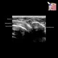
 . There is hypoechoic callus surrounding the fracture
  with echogenic mineralization more deeply. This stage of fracture healing may no longer be painful.
 at the anterior aspect of the rib close to the costochondral junction
  . Most of the callus has mineralized. There is no soft tissue swelling.