• Traumatic bleeding between the dura mater and arachnoid mater • These may be extensive – although the haemorrhage is of low pressure, the blood is unrestricted and can spread over the entire brain surface • Indirect signs: midline shift (with compression of the ipsilateral ventricle) • Some of these signs can be absent if there are bilateral collections – the frontal horns may then lie close together (with a ‘rabbit’s ears’ configuration) • The high morbidity (particularly within the elderly) is due to the associated brain swelling, contusion or laceration • Pseudomembrane: this can form around a chronic subdural haematoma • Traumatic bleeding between the cranial vault and dura mater • This is often associated with a skull fracture, which is often a fracture of the squamous part of the temporal bone (with an associated injury to the middle meningeal artery) • As the dura mater tends to adhere to the skull, the haematoma will not cross any cranial sutures but may cross a dural reflection (e.g. the falx) • The temporoparietal convexity is the commonest site (the haematoma often lies beneath a fractured squamous temporal bone) • Internal areas of low density may indicate continuing bleeding • Skull fractures: compared with vascular markings, skull fractures are straighter, more angulated, more radiolucent, and do not have corticated margins Differentiation between an extradural and subdural haematoma This includes cerebral contusions and cortical lacerations which are usually quite extensive • The injury mechanism is brain rotation with respect to the skull – it typically involves the inferior frontal lobes and the anterior temporal lobes as the sphenoid ridges and the anterior cranial fossae have irregular margins adjacent to the brain surface • ‘Contra coup’ contusion: cerebral damage lying diametrically opposite the site of impact (as defined by the skull fracture and scalp haematoma) These are less common but have a worse prognosis • The injury mechanism is the result of differential rates of rotational acceleration within the brain substance itself – this results in shearing forces damaging the axons and microvasculature • One may have to rely on so-called ‘marker’ lesions – these represent small multifocal areas of microvascular damage (with haemorrhage or infarction) and are a reliable guide to the presence of DAI but not its extent • Characteristic sites: the high parasagittal cerebral white matter
Brain trauma, degenerative disorders and epilepsy
HEAD INJURY
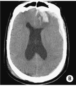
SUBDURAL HAEMORRHAGE (SDH)
DEFINITION
 it usually arises from rupture of the veins crossing the subdural space (vault fractures are an uncommon cause)
  often associated with brain damage
RADIOLOGICAL FEATURES
CT (chronic bleed)
 contralateral ventricular enlargement
  effacement of the cerebral sulci
  ‘buckling’: medial displacement of the junction between the white and grey matter
PEARLS
 dilatation of the contralateral ventricle is a bad prognostic sign
 it may show marked contrast enhancement or haemosiderin staining
EXTRADURAL (EPIDURAL) HAEMORRHAGE (EDH)
DEFINITION
RADIOLOGICAL FEATURES
CT
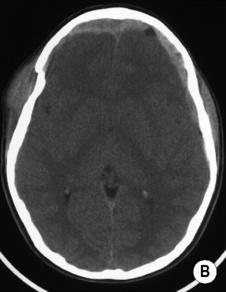
 the underlying brain is displaced but often appears intrinsically normal
PEARL
Extradural haematoma
Subdural haematoma
Location
Between the skull and dura mater
Between the dura and arachnoid mater
Cause
Trauma (fracture)
Tear of cortical bridging veins
Acute shape
Lenticular biconvex
Crescentic concave
Chronic shape
Crescentic
Elliptical
Crosses suture lines
No
Yes
Crosses a dural reflection
Yes
No
PRIMARY AND SECONDARY CEREBRAL INJURY
PRIMARY CEREBRAL INJURY
Superficial primary cerebral damage
Definition
Deep primary cerebral damage
Definition
 they occur more commonly in high-speed accidents
 the corona radiata
  the posterior corpus callosum
  the subcortical white matter


 under the temporal and occipital lobes
  along the falx cerebri


 fluid-fluid levels may be seen (denser blood elements within the dependent regions are due to acute on chronic haemorrhage)
 repeated bleeding produces variable changes in signal intensity

 risk of post-traumatic epilepsy
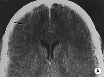
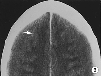
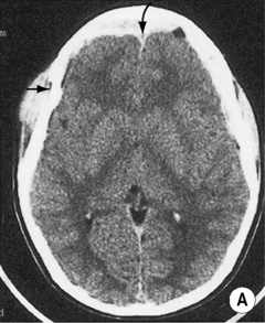
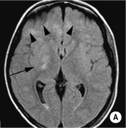
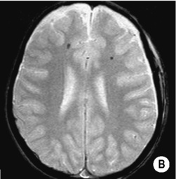
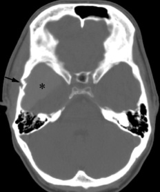
 the cortex appears abnormally thick. On close inspection, both sections show cortical vessels (arrows) displaced away from the cranial vault.*
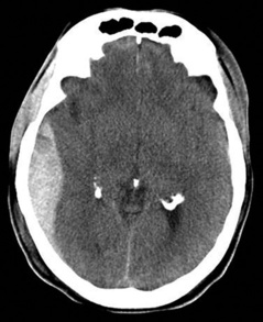
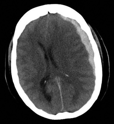
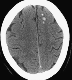
 CT. Axial ‘brain and bone windows’ demonstrate the right temporal bone depressed several millimetres with an evident fracture line (arrow) anteriorly. Soft tissue contusion overlying the fracture is also noted. A large extradural collection (curved upper arrow) anteriorly crosses the midline and displaces the falx posteriorly, and there is a second contiguous left frontal extradural collection as well. Note the generalized cerebral swelling with complete effacement of the frontal horns.*
 there can be superficial low-density areas with a mild-to-moderate mass effect – these tend to increase in the initial period and subsequently contract into a region of focal atrophy (± cavitation)
  small hyperdense haemorrhages can be present within the early stages
 chronic phase: contraction to regions of persistent (and mainly cortical) cerebral damage
 there may be hypodense foci (oedema) or hyperdense foci (petechial haemorrhages)
 it will demonstrate small dark patches of haemosiderin with characteristically normal surrounding brain
 they result from a more severe vascular component of the shearing injury and are associated with loss of consciousness at the time of injury
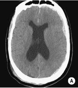
 it is caused by an increased cerebral blood volume as a result of abnormal cerebrovascular autoregulation and is a potent cause of raised intracranial pressure
 there may be effacement of the sulci and cisterns with loss of the grey/white matter interface