Fig. 19.1
TAAA preoperative image
MR angiography with contrast enhancement can be used for aneurysm diagnostics, procedural planning as well as follow-up, but it is often more cumbersome and time consuming to achieve a satisfactory result.
Diagnostic angiography has mostly been replaced by CT scans in this patient category since angiography is invasive and places an extra contrast burden on the patient. In selected cases, angiography is used as an additional imaging tool, often in conjunction with embolization of branching vessels which might be performed prior to the aortic reconstruction
6 Management
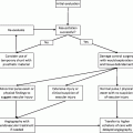
Indications for Treatment
1.
Symptomatic disease
(a)
Abdominal pain and tenderness with confirmation by imaging.
(b)
Ruptured aneurysm (open repair).
2.
Asymptomatic disease
(a)
Rapidly expanding aneurysm (≥5 mm/year).
(b)
Maximal aneurysm diameter >55–60 mm.
6.1 Open Treatment
Open repair is commonly the choice in symptomatic or ruptured aneurysms or in trauma patients due to the need for extensive customization of the graft when performing an endovascular repair.
1.
Transabdominal repair: Suprarenal aneurysm requiring a supraceliac or supra SMA crossclamp.
2.
Thoracoabdominal repair: For Crawford type 1–4 extent disease. Often performed with left heart bypass, visceral perfusion, and sequential crossclamping of the aorta.
3.
Hybrid repair: In high-risk patients not suitable for endografting due to anatomical restrictions. Extraanatomic visceral artery bypass (from iliacs or aorta) combined with endovascular stentgrafting of the aorta.
6.2 Endovascular Treatment
6.2.1 Stent Graft Design (Fig. 19.2)
Fig. 19.2
TAAA graft. Image courtesy of COOK Medical Inc
The implanted devices are constructed using the AAA Zenith endograft platform by Cook (Cook Medical, Bloomington, IN, USA) comprising stainless steel Z stents and woven polyester fabric. The key difference between standard infrarenal EVAR and branched stent grafts is the presence of a reducing stent and side branches. The reducing stent, which is located distal to the sealing zone, converts the main body into a 16, 18, or 22-mm cuff bearing segment. This diameter is planned based on the aortic lumen so that it can accommodate both the main body and the bridging covered stents between the graft side branches and the visceral vessels. Aortic sidebranches, as opposed to fenestrations, are used when there is not expected to be any contact between the main body of the endograft and the aorta at the level of the visceral arteries. The diameter of the distal segment is adjusted to be sealed either to the diameter of a previous graft, in those patients who have already undergone open or endovascular infrarenal aneurysm repair or return to a diameter of 22 mm, which will serve as a standard diameter for the overlap of a distal 24-mm bifurcated endograft that is placed simultaneuously. Depending on the proximal extent of the aneurysm, additional endografts may be used proximally. The proximal sealing zone of the graft consists of one or two sealing stents with or without an uncovered proximal stent. Proximal barbs are added to the first sealing stent or to the uncovered stent for fixation to the aortic wall.
Related posts:

Stay updated, free articles. Join our Telegram channel

Full access? Get Clinical Tree


