Breast is a modified sweat gland.
Functional unit of breast is terminal ductulo lobular unit (TDLU); site of origin of most breast pathologies.
Three zones from superficial to deep (Figure 38.1):
Premammary zone—Subcutaneous zone
Mammary zone—Contains most of the lobar ducts, TDLUs, and fibrous stromal elements of the breast
Retromammary zone—Contains fat, blood vessels, and lymphatics
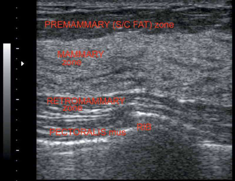
Figure 38.1 Illustrates normal zones of breast.
Echogenicity of structure from superficial to deep:
Hyperechoic—Skin (<2 millimeters thick)
Hypoechoic—Subcutaneous fat (Premammary s/c fat—Lobulated and more hyperechoic than fat elsewhere)
Hyperechoic—Fibroglandular parenchyma (12–20 ducts along with their lobules, fat, and stroma constitutes the breast parenchyma
Hypoechoic—Retromammary fat
Hyperechoic—Muscle (Pectoralis major)
Echogenicities of various structures:
Hyperechoic structures—Compact interlobular stromal fibrous tissue, anterior and posterior mammary fascia, Cooper’s ligament (thin echogenic bands), duct wall, and skin.
Isoechoic—Loose intralobular and periductal stromal fibrous tissue, fat, and epithelial tissues in ducts and lobules.
Hypoechoic—Retromammary fat.
Advantages and indications
1. Ideal in young, pregnant, and lactating females (nonionizing)
2. To differentiate cystic versus solid lesions
3. In tender/inflamed breasts (no compression required as in mammography)
4. To differentiate benign versus malignant lump
5. Follow-up of cysts
6. For lymph nodes
7. As a guide for interventional procedures
8. For implants
9. For clinical breast mass with indeterminate mammogram
10. Follow-up of cancer patients on chemotherapy
11. Suspicious lump in males
Limitations
1. Operator dependent.
2. Low sensitivity especially for microcalcifications.
Technique—Scanning is done in supine and contralateral oblique position with arms comfortably under the head.
Side—Right (R) or Left (L)
The clock face with the center at the nipple (1–12 o’clock position)
Zones—Nipple (N), subareolar (SA), axillary (AX), and three circular concentric zones outside the subareolar zone (one, two, and three)
Probe orientation—Radial (RAD), antiradial (ARAD), horizontal, vertical, and oblique planes
Depth of the lesion from the skin
Characteristics to be noted: (Table 38.1)
1. Shape—Round, oval, and its extension along the ducts
2. Size, both in short and long axes, to look for interval changes, if any, on follow-up scans
3. Surface—Smooth, irregular, lobulated, and spiculations
4. Echotexture
Hypo/iso/hyperechoic
Homo/heterogeneous
Cystic/calcific component, if any
5. Fixity to surrounding tissues and underlying muscles
6. Doppler findings
Table 38.1 Illustrating differentiating features of benign and malignant breast lesions
Related posts:
Stay updated, free articles. Join our Telegram channel

Full access? Get Clinical Tree


