Imaging Methods
Breast imaging generally refers to mammography. Mammography can detect a significant number of tumors not found by palpation or self-breast examination. Even if a mass is palpable, there are no reliable physical characteristics to distinguish benign from malignant lesions. The U.S. Preventive Services Task Force (USPSTF) has concluded that evidence is insufficient to assess the benefits and harms of a clinical breast examination in women 40 years or older. The primary purpose of mammography is to detect small breast cancers and, by so doing, to improve outcomes. In young women, the breast is often extremely dense. The density of normal fibroglandular tissue is the same as the density of a carcinoma. In young women, not only is the incidence of breast cancer low, but it is also very difficult to tell whether a cancer is present amid the normal parenchyma. As women age, fatty replacement of the breast and atrophy of the parenchyma occur. Because fat is lucent (dark) on a mammogram and cancer is dense (white), tumors are more easily visualized as a woman ages. The amount of dense fibroglandular tissue is partly due to hormonal stimulation. In older women receiving estrogen replacement therapy, the density of the breast tissue increases, making tumors more difficult to see ( Fig. 4.1 ). The density of the breast also increases during pregnancy and lactation; however, screening and evaluation of pregnancy-associated breast cancer with digital mammography or tomography is usually appropriate.

Mammograms are done for two distinct purposes: (1) screening and (2) workup of an abnormality. Standard mammogram images are like other plain x-rays in that they are two-dimensional (2D) images of three-dimensional (3D) structures. Projections are usually obtained in what are referred to as the craniocaudal (CC; superior to inferior) and medial lateral oblique (MLO) views. The latter, a somewhat angled lateral view, allows better visualization of the tail of the breast tissue as it extends out toward the axilla than is possible on a true lateral view. On the CC view, it is often not easy to tell which is the medial and which is the lateral aspect. By convention, the identifying markers or technologist’s initials are placed along the lateral edge of the breast. Workup mammograms have special views and often use magnified images. At the present time, all mammograms are done using full-field digital mammography (FFDM) receptors and not film. This has allowed a radiation dose reduction of about 25%, as well as having immediate images to view.
In the last decade, there has been increasing use of a technique known as digital breast tomosynthesis (DBT). With this technique, the x-ray tube rotates over a 15- to 50-degree arc and allows computer manipulation of the dataset to create images in several planes of the breast thickness. Objects not in the plane selected are blurred out. Breast doses are reasonably comparable from either a two-view FFDM or one-view DBT. Some centers use a full-field digital mammogram combined with a one-view tomosynthesis (doubling the normal breast dose), and others may use a combined full-field digital mammogram and two-view tomosynthesis. Recently, the situation has become more complicated as it is possible to use the same 3D tomosynthesis dataset to compute a synthetic 2D image, obviating the need for the standard full-field digital mammogram. Tomosynthesis has been shown to increase cancer detection rate slightly above that for a screening mammogram (from about 6/1000 to about 7/1000). One main value of tomosynthesis during screening is that there can be rapid resolution of apparent abnormalities caused by the superimposition of normal structures, which obviates the need to call back many patients for a workup mammogram.
Ultrasound examination is often used as an adjunct to mammography to determine whether a lesion is solid or cystic and to localize the lesion for needle biopsy. Ultrasound can be a useful method in conjunction with diagnostic mammogram, but should not be relied on as a screening method for breast cancer. Computed tomography scanning is not indicated for examination of the breast. There are nuclear medicine techniques to image breast lesions (breast-specific gamma imaging and positron emission mammography), but their radiation dose is 15 to 30 times higher than from a digital mammogram, and their use for screening is not indicated. Magnetic resonance imaging (MRI) is occasionally used in select patients, including for screening of women with more than a 20% lifetime probability of breast cancer, or for problem solving.
Interpretation and Workup
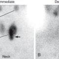
Great variation is often found among women in the appearance of their breast tissue. Fortunately, most women have symmetric tissue when one breast is compared with the other. Any asymmetries in density should be examined carefully because they may represent a cancer ( Fig. 4.2 ). In addition to asymmetric masses, another sign of breast cancer is tiny grouped calcifications. Often called microcalcifications , these are usually fine (approximately ≤1 mm), linear, and with pleomorphic, or branching morphology. Most women, as they age, have benign calcifications within the breast. These are usually rounded calcifications greater than 2 mm in diameter ( Fig. 4.3 ). In women over the age of 60 years, serpiginous calcifications can normally be seen within the blood vessel walls. Associated indirect signs of malignancy also may be present. These include focal skin thickening or dimpling due to an underlying tumor, unilateral nipple retraction, and vascular asymmetry (increased vascularity due to the tumor).


Related posts:
Stay updated, free articles. Join our Telegram channel

Full access? Get Clinical Tree


