• The breast lies on the chest wall and on the deep pectoral fascia • High spatial resolution is required to detect microcalcification: a short exposure time limits any movement artefact • There is a narrow range of inherent breast densities (as it is predominantly fatty tissue): low molybdenum energy peaks (17.5 and 19.6keV) provide high contrast (filtering reduces extraneous radiation) • Breast compression: this reduces geometric and movement unsharpness • Mediolateral oblique (MLO) view (standard): • Craniocaudal (CC) view (standard): • Paddle views (supplementary): • True lateral view (supplementary): • Magnification views (supplementary): • Eklund technique (supplementary): • Exaggeration of the normal cyclical proliferation and involution of breast tissue with the development of fibrosis • A benign tumour arising from the TDLU (fibrous stroma + epithelial ductal structures) • Requires biopsy for diagnosis unless <25 years old (to exclude malignancy) • Juvenile fibroadenoma: a more cellular variant occurring at a younger age • Phyllodes tumour: a fibroepithelial tumour similar to a giant fibroadenoma, affecting an older age group
Breast
NORMAL ANATOMY
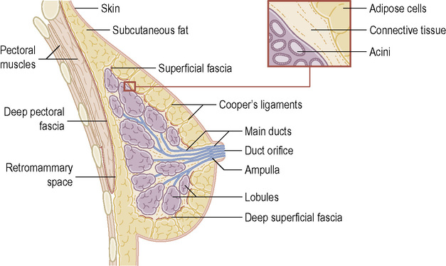
 the superficial pectoral fascia envelops the breast
  suspensory ligaments (Cooper’s ligaments) connect the two layers
2 components
METHODS OF IMAGING
Imaging in mammography
 it requires a very small focal spot (0.1–0.3mm)
  grids are used to reduce scatter and increase contrast
  digital mammography is now used
 it improves contrast (it reduces scatter)
  it reduces radiation dose (less tissue needs to be penetrated)
  it achieves uniform image density
  it separates superimposed breast tissues
  it highlights rigid tumours (glandular tissue is compressible)
Mammography (digital/analogue) (MMG)
 Evaluation of breast symptoms and signs, including masses, skin thickening, deformity, nipple retraction, nipple discharge and nipple eczema
 Follow-up of breast cancer patients
 Guidance for biopsy or localization of lesions not visible on ultrasound
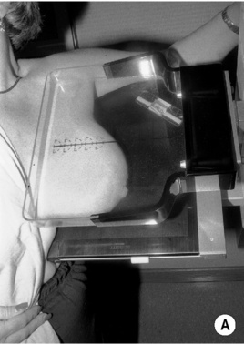
 The XR beam is directed from superomedial to inferolateral (usually at 30–60°)
  compression is applied obliquely across the chest wall and perpendicular to the long-axis pectoralis major
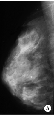
 The only view demonstrating the entire breast tissue on a single image
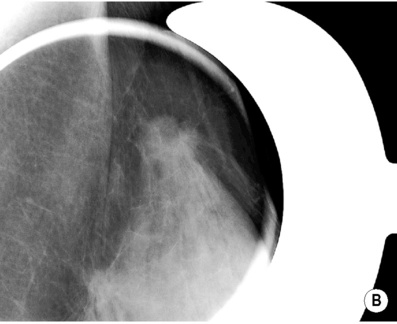
 The XR beam travels from superior to inferior
  the breast is pulled forward and away from the chest wall with compression applied from above
INTRODUCTION
FIBROCYSTIC CHANGE
DEFINITION
 regression with pregnancy and menopause
  increased risk for developing certain types of cancer (e.g. ductal carcinoma in situ (DCIS))
BENIGN MASS LESIONS
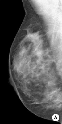
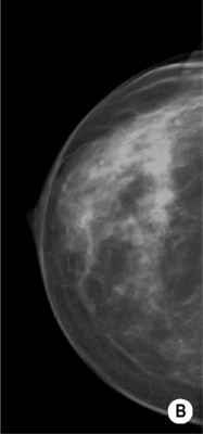
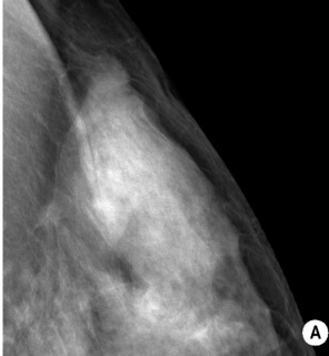
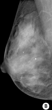
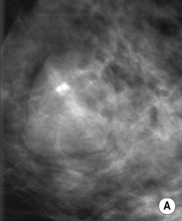
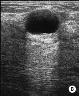
FIBROADENOMA
DEFINITION
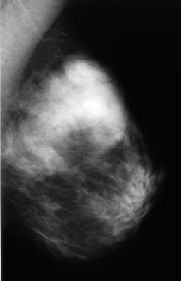
 it often enlarges during pregnancy and regresses after the menopause
  it is the most common cause of a benign solid mass in the breast
RADIOLOGICAL FEATURES
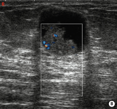
US
 < 25% are locally aggressive requiring clear surgical margins
  large fibroadenomas or those rapidly increasing in size are excised to avoid missing a phyllodes tumour
Breast


 ducts open onto the skin surface (and are seen as small raised nodular structures called Morgagni’s tubercles)


 this is usually replaced by fatty tissues in older women with loss of the lobular units


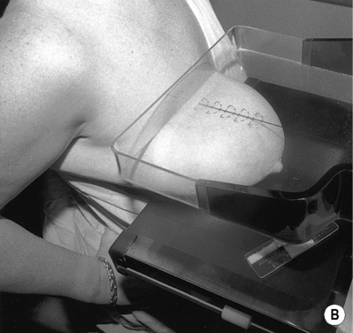
 the nipple is in profile
  the nipple is positioned at the level of the lower border of the pectoralis major, with the muscle across the posterior border of the film at 25–30° to the vertical
 it demonstrates virtually all of the medial tissue and the majority of the lateral tissue (with exclusion of the axillary tail of the breast)
  the depth of breast tissue should be < 1cm of the distance from the nipple to the pectoralis major on the MLO projection

 it defines the margins of a mass

 it increases the accuracy of wire localizations of non-palpable lesions


 T2WI: diffuse high SI (2nd half of cycle): T1WI + Gad: patchy enhancement
 diffusely scattered calcifications
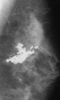
 a ‘teacup’ configuration of calcium within the cystic spaces
 common between 20 and 50 years old (peak between 40 and 50 years old)
 perform cytology only if there are suspicious imaging features or if the aspirate is bloodstained
 coarse mural curvilinear calcification
 they can occur at any age and are often palpable
 on histology it may be reported as normal breast tissue
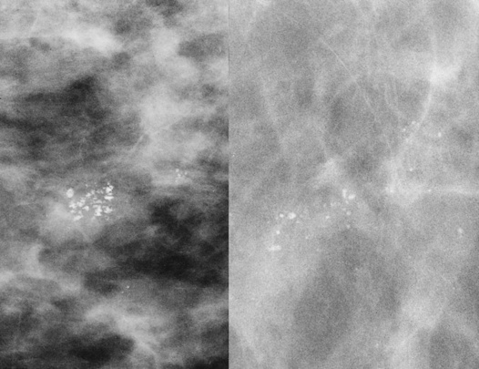

 peak incidence – 3rd decade
  multiple in 10–20%
 coarse calcifications may develop (particularly in older women)
 isoechoic or mildly hypoechoic relative to fat ± a thin echogenic pseudocapsule
  a surrounding halo (Mach effect)
  displaced normal vessels around the edge of the lesion


