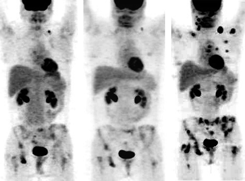
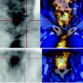
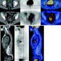
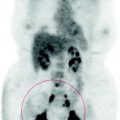
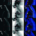
Fig. 51.1
CT scan shows multiple bone thickening lesions. The PET characterizes the lesions with a high metabolism of glucose. This information must be critically assessed
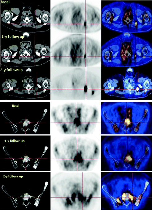
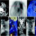
Fig. 51.2
The PET-CT scan shows a hypodense lymph node in the left clavicular fossa, with volumetric and metabolic activity progression over time. The follow up at two years shows the appearance of a new dorsal—retroscapular node. In a similar way, it detects progression of the bone disease
