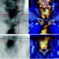
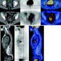
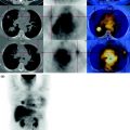
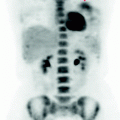
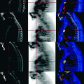
Fig. 49.1
MIP image: slight increase in the concentration of FDG in the left axilla with small nodes characterized by limited metabolism, therefore deemed as reactive (yellow arrow). The liver shows physiological glucose consumption and presents no metastatic disease. Slight focal nodular uptake in the left buttock due to a granuloma (red arrow)
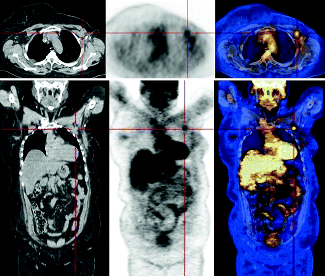
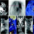
Fig. 49.2


The CT scan shows the previous left mastectomy with axillary dissection. Adjacent to the metal clips thickening of the pectoral muscle is evident and an axillary centimetric lymph node that shows a slight increase of glucose metabolism at the PET scan
Stay updated, free articles. Join our Telegram channel

Full access? Get Clinical Tree


