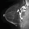
Fig. 6.1
Sagittal T1-weighted fat-saturated post-contrast maximum intensity projection (MIP) image (a), sagittal T2-weighted fat-saturated image (b), sagittal T1-weighted fat-saturated image (c), and axial T1-weighted fat-saturated image (d)
6.2 Saline Rupture
Teaching Points
Implant rupture is a well-known complication and is the main cause of implant removal. Most ruptures have no obvious traumatic origin, and rupture is sometimes present in asymptomatic patients. MRI is generally accepted as the technique of choice for evaluating the integrity of silicone implants, with a sensitivity of 70–94 % and a specificity of 85–100 %. Patients with saline implant rupture usually report an obvious change in size with fluid extrusion, for which breast MRI is not necessary for diagnosis. Saline implants follow fluid signal on all sequences and have a valve to allow volume adjustment, which helps to identify them on MRI. In our case example (Figs. 6.1 and 6.2), saline implant rupture with complete collapse was incidentally found on a high-risk screening breast MRI.
Image Findings
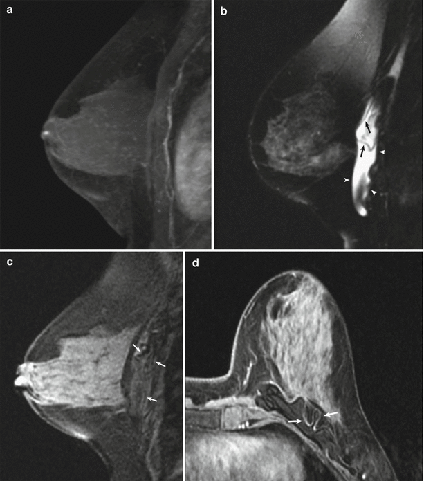
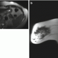
Fig. 6.2
Saline implant rupture. (a) Sagittal T1-weighted post-contrast MIP demonstrates no suspicious enhancement. (b) Sagittal T2-weighted fat-saturated image demonstrates a collapsed saline implant (arrows) with surrounding T2 hyperintensity (arrowheads). Sagittal (c) and axial (d) T1-weighted fat-saturated images demonstrate corresponding T1 hypointensity surrounding the collapsed implant (arrows)
6.3 History
32-year-old woman status post motor vehicle accident. History of implant (Fig. 6.3).
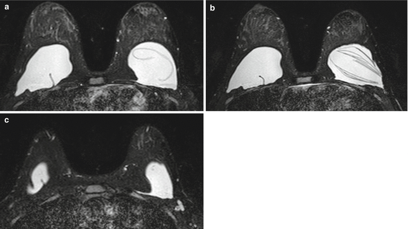
Fig. 6.3
(a–c) Axial T2-weighted fat-saturated MR images through both breasts
6.4 Silicone Implant Rupture 1
Teaching Points
MR imaging can diagnose silicone breast implant rupture with a high sensitivity and specificity, with a positive predictive value of about 99 %. Silicone implant rupture is usually unnoticed by the patient because free silicone is kept in place by the surrounding fibrous capsule, which begins forming immediately after surgery as a natural foreign body reaction.
Extracapsular silicone implant rupture is defined as rupture of both the implant shell and the fibrous capsule, with macroscopic silicone leakage extending beyond the fibrous capsule into surrounding tissues and rarely to distant body regions (Fig. 6.4). Intracapsular rupture is defined as rupture of the implant shell with an intact fibrous capsule. The “linguine sign” (the presence of multiple curvilinear, low–signal-intensity lines within the silicone implant) is considered the most reliable MRI characteristic for intracapsular rupture and is often seen with extracapsular implant ruptures.
Radial folds, which are considered to be normal variants, are infoldings of the implant membrane into the silicone implant. On MRI, they appear as blind-ending lines extending from the surface of the implant (Fig. 6.4c).
Image Findings

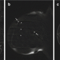
Fig. 6.4


Left extracapsular silicone implant rupture and right silicone implant radial fold. (a, b) Selected axial T2-weighted fat-saturated images demonstrate collapse of the left silicone implant shell (white arrows) with extravasation of silicone into the fibrous capsule. The “linguini sign” (black arrowheads in b) of intracapsular rupture is evident. Complex radial folds (black arrow in b) are present involving the right silicone implant shell without evidence of right implant rupture. (c) On a more inferior T2-weighted fat-saturated axial image, there is extracapsular leak of silicone (white arrows) laterally into the soft tissue along the left chest wall
Related posts:
Stay updated, free articles. Join our Telegram channel
Full access? Get Clinical Tree
