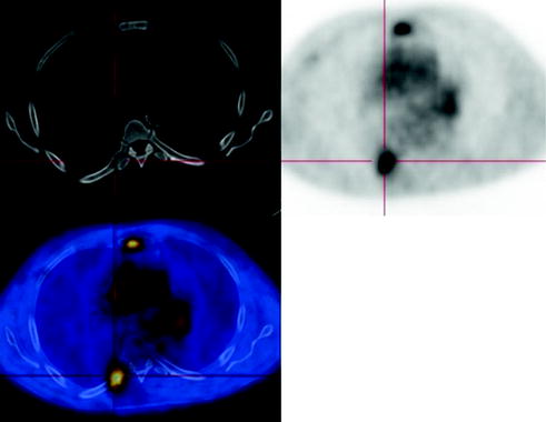
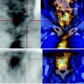
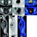
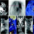
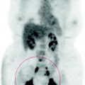
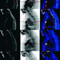
Fig. 70.1
MIP image: bone metastases have a metabolic activity higher than that of the primary tumor, that probably has a contextual internal colliquation
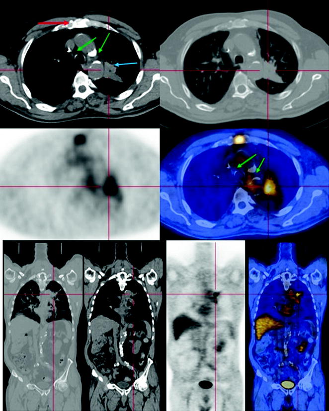
Fig. 70.2
The PET-CT scan shows a solid mass characterized by pathological metabolism at the posterior segment of the left upper lobe. This mass is spiculated, infiltrating the surrounding tissues, is inseparable from the mediastinal vascular structures and determines modest atelectasis of the parenchyma. The body of the sternum presents a lytic lesion with high metabolism. At the aortopulmonary window and the Barety space, CT shows some subcentimetric lymph nodes that do not have abnormal glucose consumption
