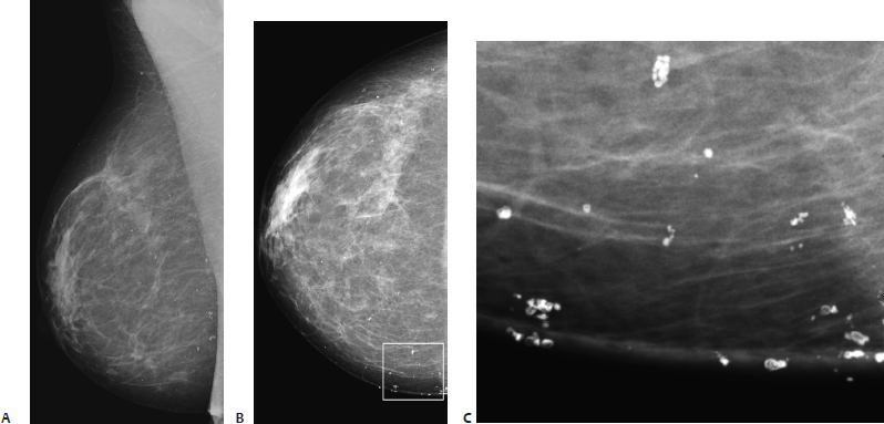
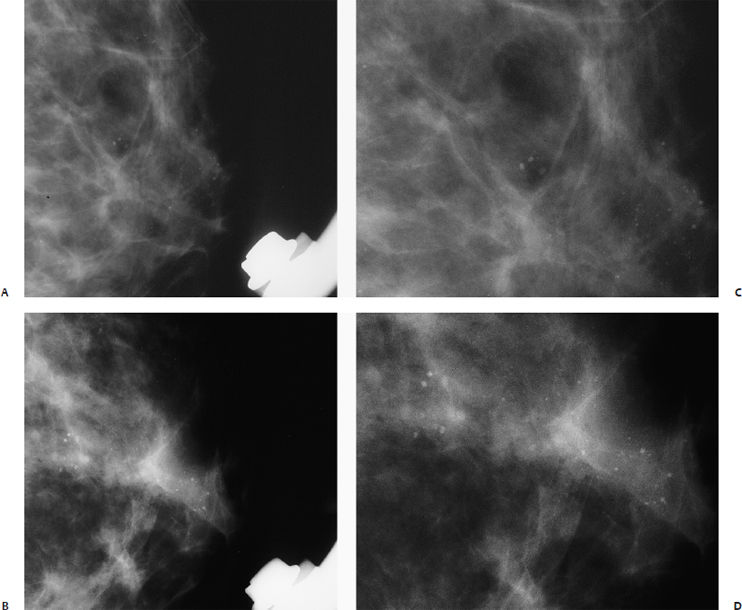
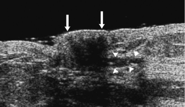
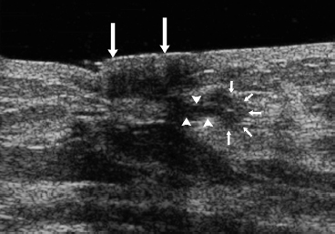
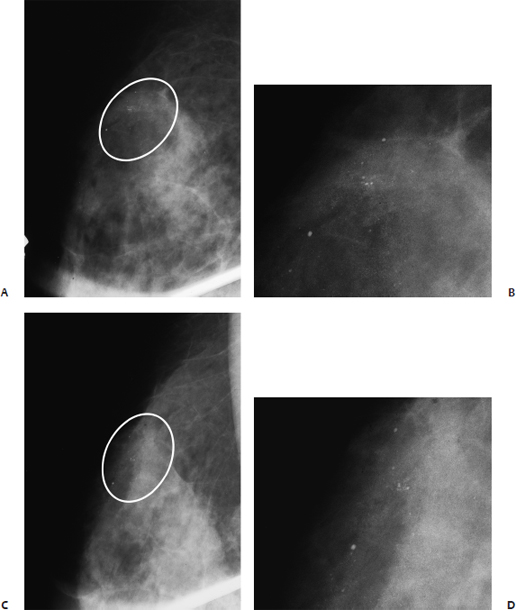
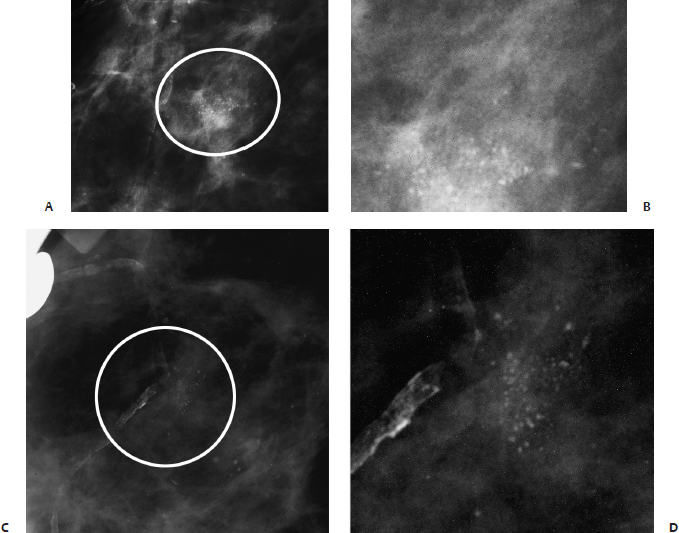
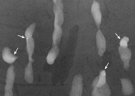
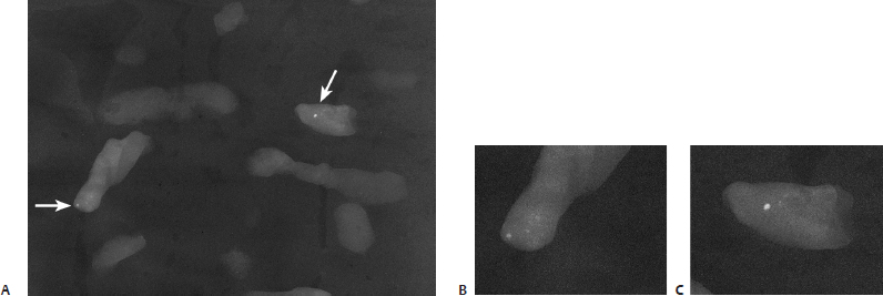
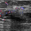
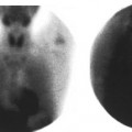
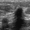
16 Calcifications: Small Round/Punctate A 41-year-old woman presents for screening mammogram. • Normal exam Calcifications (Fig. 16.1) • Type: skin calcifications • Distribution: scattered, diffuse Fig. 16.1 There are multiple scattered round and polygonal skin calcifications, many with lucent centers. (A) Right MLO mammogram. (B) Right CC mammogram. (C) Enlargement of medial CC mammogram, which is marked by a square on B. • BI-RADS assessment category 2, benign finding • Skin calcifications are commonly in the medial side of the breast. They are well defined, round or polygonal in shape, with lucent centers. If they are superimposed upon the breast parenchyma, they may be mistaken for intraparenchymal calcifications. Tangential views will distinguish skin calcifications from intraparenchymal ones. This case illustrates that digital mammography is superior to film screen technique in displaying these calcifications, because the skin and subcutaneous tissues are better visualized with the digital technique. • These calcifications are within the sebaceous glands and may be the result of chronic folliculitis. Kopans DB, Meyer JE, Homer MJ, Grabbe J. Dermal deposits mistaken for breast calcifications. Radiology 1983;149:592–594 A 49-year-old woman presents with new left subareolar calcifications on her screening mammogram. • Normal exam Calcifications (Figs. 16.2 and 16.3) • Type: punctate • Distribution: regional Fig. 16.2 Numerous round, punctate calcifications are present in the subareolar region. (A) Left MLO magnification mammogram. (B) Enlargement of calcifications in (A). (C) Left CC magnification mammogram. (D) Enlargement of calcifications in (C). Fig. 16.3 Specimen needle core biopsy radiograph. The round, punctate calcifications (arrows) are easily identified within this biopsy specimen. Frequency • 10 to 13 MHz (Figs. 16.4 and 16.5) Fig. 16.4 Left radial breast sonogram. Under the nipple (large arrows), there were several main ducts (arrowheads). Fig. 16.5 Left radial breast sonogram. The main ducts (arrowheads) led to lobulated structures (small arrows), consistent with lobular hyperplasia. Nipple (large arrows). • Cysts; besides calcifications within microcysts, atypical lobular hyperplasia was present. • BI-RADS assessment category 3, probably benign; short-interval follow-up • The calcifications in this case are round because they are within microcysts. This atypical lobular hyperplasia was inadvertently adjacent to these microcysts. • High-frequency sonography is able to delineate ductal and lobular details that were not visible with lower-resolution equipment. Gershon-Cohen J, Berger SM, Curcio BM. Breast cancer with microcalcifications: diagnostic difficulties. Radiology 1966;87:613–622 Teboul M, Halliwell M. Atlas of Ultrasound and Ductal Echography of the Breast. 2nd ed. Cambridge: Blackwell Science; 1996:154–201 A 45-year-old woman presents with right breast calcifications that have increased in number since her prior screening mammogram performed 2 years ago. • Normal exam Calcifications (Figs. 16.6 and 16.7) • Type: punctate • Distribution: diffuse/scattered Fig. 16.6 In the upper outer quadrant, there are scattered round, punctate calcifications (circle). (A) Right MLO magnification view. (B) Enlargement of calcifications in A. (C) Right accentuated CC magnification view. Fig. 16.7 (A) Specimen radiograph of breast tissue cores from percutaneous biopsy. The punctate calcifications in the specimen (arrows) mirror the appearance of the punctate calcifications in Fig. 16.6. (B) and (C) Enlargements of calcifications in (A). • Cysts; benign fibrous and cystic changes with calcifications in cysts • BI-RADS assessment category 2, benign finding • Scattered punctate or round microcalcifications are produced by a variety of benign conditions, including cystic hyperplasia, papillomatosis, sclerosing adenosis, adenosis, and atypical lobular hyperplasia. Gershon-Cohen J, Berger SM, Curcio BM. Breast cancer with microcalcifications: diagnostic difficulties. Radiology 1966;87:613–622 A 57-year-old woman presents with increasing right breast calcifications. • Normal exam Calcifications (Fig. 16.8) • Type: punctate • Distribution: grouped/clustered Fig. 16.8 In the upper outer quadrant of the right breast, there is a cluster of punctate calcifications (circle). (A) Right MLO magnification mammogram. (B) Enlargement of calcifications in A. (C) Right CC magnification mammogram. (D) Enlargement of calcifications in C.
Case 16.1: Skin
Case History
Physical Examination
Mammogram
Management
Pearls and Pitfalls
Suggested Reading
Case 16.2: Fibrocystic Change
Case History
Physical Examination
Mammogram
Ultrasound
Pathology
Management
Pearls and Pitfalls
Suggested Reading
Case 16.3: Fibrocystic Change
Case History
Physical Examination
Mammogram
Pathology
Management
Pearls and Pitfalls
Suggested Reading
Case 16.4: Fibrocystic Change
Case History
Physical Examination
Mammogram
Related posts:


Stay updated, free articles. Join our Telegram channel

Full access? Get Clinical Tree


