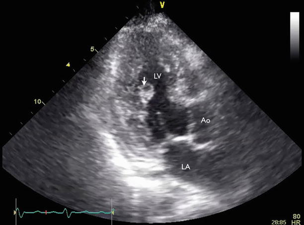
Fig. 13.1
Transthoracic echocardiography (TTE) parasternal long-axis view showed a 3.7 × 3.6-cm, highly mobile echodensity (arrows) with frond-like projections in the left ventricle (LV), attached to the posterior mitral valve leaflet and subvalvular apparatus. Ao aorta, LA left atrium
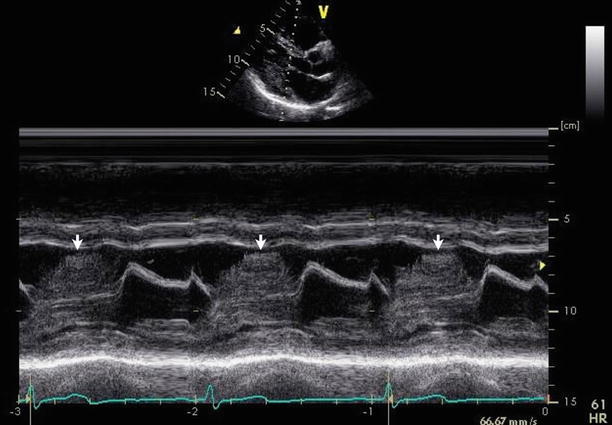
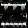
Fig. 13.2
M-mode echocardiography at the mitral valve showed the presence of an echodensity intermittently protruding into the left ventricular outflow tract during systole (arrows)
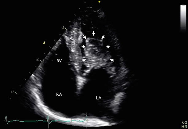
Fig. 13.3
TTE apical four-chamber view showed the mass (arrows) of the mitral valve. RA right atrium, RV right ventricle
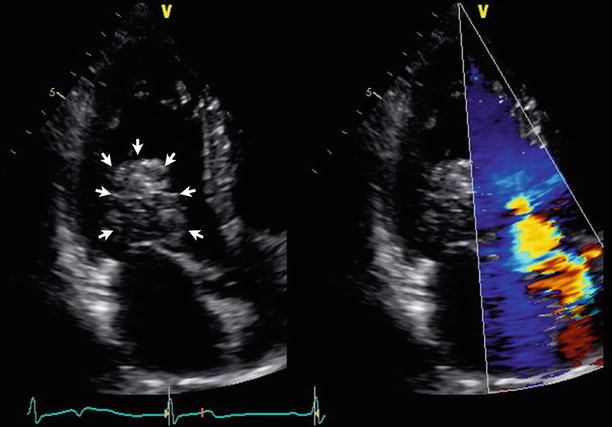
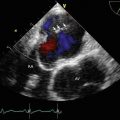
Fig. 13.4
TTE apical five-chamber view showed that the mass (arrows) protruded into the left ventricular outflow tract (LVOT) during systole. The mass also caused flow acceleration in the LVOT, corresponding to the patient’s systolic murmur
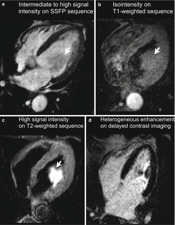
Fig. 13.5
(a–d) Cardiac MRI confirmed the findings, with tissue characteristics consistent with cardiac myxoma. SSFP steady-state free precession
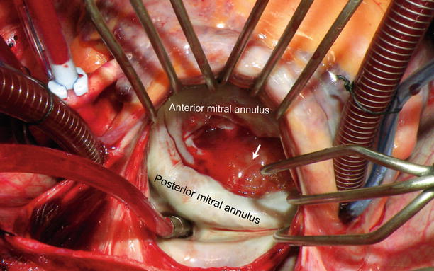
Fig. 13.6
Intraoperative findings showed that the mass (arrow) was attached to the head of the posteromedial papillary muscle and several chords going to the P2 segment of the posterior mitral valve leaflet (Courtesy of Dr. Joseph Sabik, Cleveland Clinic)
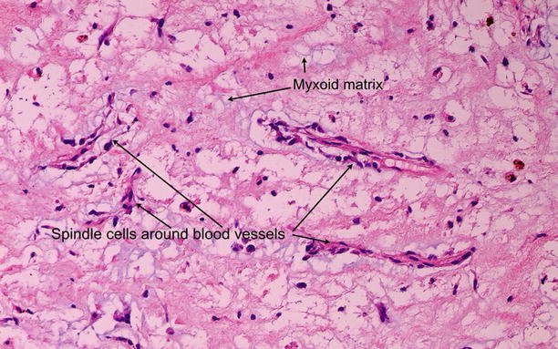
Fig. 13.7
Pathology confirmed the diagnosis of mitral valve myxoma. (Arrow indicates mass.) (Courtesy of Dr. Carmela Tan and Dr. Rene Rodriguez, Cleveland Clinic)
Video 13.1 Transthoracic echocardiography (TTE) parasternal long-axis view showed a 3.7 × 3.6-cm, highly mobile echodensity with frond-like projections in the left ventricle, attached to the posterior mitral valve leaflet and subvalvular apparatus (AVI 5351 kb)
Video 13.2 TTE apical four-chamber view showed the mass (arrows) of the mitral valve (AVI 4775 kb)
13.2 Case 2. Blood Cyst
An 84-year-old man with past medical history of hypertension and dyslipidemia was sent for echocardiographic evaluation because of near syncope.
13.2.1 Learning Points
Blood cysts are congenital in origin and most frequently have been found as a postmortem finding in fetuses and infants less than 6 months of age [5]. Histologically, they are diverticuli lined with flat, endothelium-like cells on both sides, containing bloody fluid. Echocardiographic findings include a thin-walled, well-circumscribed mass with an echolucent core (Figs. 13.8, 13.9, 13.10, and 13.11). The presence of microbubbles inside the cystic cavity during diastole (confirming the passage of blood between the ventricular cavity and the cyst lumen) has been described as a pathognomonic sign of the presence of a blood cyst [6]. The cyst is most commonly present at an atrioventricular valve; it occurs less often in semilunar valves and is also found at papillary muscle [7] or the atrialized right ventricular portion of Ebstein’s anomaly.