Carpal Tunnel Syndrome
KEY FACTS
Terminology
Imaging
 Intraneural hyperemia
 Retinacular bowing
 Flattening ratio: Width:depth ratio > 3 of median nerve ↑ with CTS
 Homogeneously hypoechoic with loss of normal fascicular pattern
 If either CSA proximal to or distal to carpal tunnel is > 14 mm² or palmar volar bowing of flexor retinaculum at outlet > 1 mm, sensitivity, specificity & accuracy are 100%, 84%, and 93%, respectively
IMAGING
General Features
Ultrasonographic Findings
 Swelling of median nerve either immediately proximal to, within, or immediately distal to carpal tunnel
 Measure CSA by tracing around nerve perimeter
 2 main measurement methods based on median nerve CSA, either of which can be used
 Median nerve CSA measured at 4 points: Immediately proximal to tunnel inlet, at tunnel inlet, at tunnel outlet, & distal to carpal tunnel
 Optimal cutoff point: Median nerve CSA ≥ 14 mm²
 Median nerve CSA ≥ 14 mm² at proximal or distal to carpal tunnel: Strong positive predictor of CTS (sensitivity/specificity/accuracy > 88%)
 Median nerve CSA measured at proximal 1/3 of pronator quadratus muscle level (CSAf) & immediately proximal to tunnel inlet, at tunnel inlet, & at tunnel outlet (CSAc)
 Difference between pronator quadratus level measurement (CSAf) & any carpal tunnel measurement (CSAc) should not be > 4 mm²
 Difference between CSAc & CSAf (ΔCSA) of > 4 mm² is strong positive predictor of CTS


 deep to the flexor retinaculum
  & superficial to the flexor tendons. The nerve usually divides just beyond the tunnel outlet.
 lies immediately below the flexor retinaculum
  & superficial to the flexor digitorum tendons, which are surrounded by the ulnar bursa
  & the flexor pollicis longus, which is surrounded by the radial bursa
  . The extrinsic ligaments
  lie above the carpal bones.
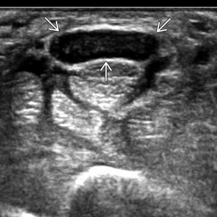
 . A nerve cross-sectional area (CSA) > 14 mm² is a positive predictor for CTS. Note also the loss of neural fascicular structure, which is an additional sign of CTS.