and Gerald Wyse1
(1)
Department of Radiology, Cork University Hospital, Cork, Ireland
Abstract
A full clinical history, physical examination, and review of the study indication should be performed prior to every cerebral angiogram. Perform noninvasive imaging initially with magnetic resonance (MR), computed tomography (CT), and/or CT/MR angiography. Closely review all imaging and laboratory data prior to invasive angiography.
Keywords
Diagnostic cerebral angiographyGroin accessClosureMRCTHistory and physicalDiagnostic Evaluation
A full clinical history, physical examination, and review of the study indication should be performed prior to every cerebral angiogram. Perform noninvasive imaging initially with magnetic resonance (MR), computed tomography (CT), and/or CT/MR angiography. Closely review all imaging and laboratory data prior to invasive angiography.
Indications for Performing a Diagnostic Cerebral Angiogram
Despite the advances of CT and MR angiography, invasive diagnostic cerebral angiography still has a broad number of indications. Today cerebral angiograms are commonly performed to access dynamic process such as AV shunts or following intracranial interventions such as coil embolization of aneurysms. Cerebral angiography can be performed to further investigate patients with:
Stenosis or occlusion
Aneurysm
Vascular malformations
Vasculitis
Vascular tumors
Previous intracranial intervention
Contraindications
Absolute
History of anaphylaxis to iodinated contrast
Patient informed refusal
Relative
Coagulopathy
Contrast allergy
Abnormal renal function
Uncontrolled hypertension
Decompensated heart failure
Pregnancy
Patient Preparation
Nil by mouth for 6–8 h preferable
Peripheral IV access
Correct coagulopathy and other contraindications where possible
Relevant Aberrant Anatomy
Familiarity with common vascular anatomy is essential. Understanding of the common aberrant or variant anatomy is critical to avoid misinterpretation.
Common Arch Variants
“Normal” (70 %).
Common left common carotid and innominate origin (termed “bovine” arch) (13 %).
Left common carotid arises from the innominate (9 %) (also termed “bovine” arch).
Left vertebral artery directly from arch (5 %).
Left brachiocephalic trunk (2.7 %).
Aberrant right subclavian (0.5 %).
Common Intracranial Variants
Complete circle of Willis (20–25 %)
Hypoplasia of one or both posterior communicating arteries (PComs) (34 %)
Hypoplastic/absent A1 segment of the anterior cerebral artery (ACA)
Origin of posterior cerebral artery (PCA) from the internal carotid artery (ICA) with and absent or hypoplastic P1 segment (17 %) (fetal PCom or fetal circulation)
Infundibular dilatation of the PCom origin (10 %)
Preprocedure Medications
Sedation of the patient is not routinely required. An oversedated patient can be difficult to neurologically assess, and an acute complication may be missed. In addition noncompliance from a sedated patient may lead to a poor quality or even nondiagnostic study.
Procedure
Access
A common femoral artery (CFA) groin approach, the right groin in particular, is favored for access. The femoral artery is easily compressed post procedure and is associated with a low puncture site complication rate even in the setting of antiplatelet agents.
Radial access continues to grow in popularity for coronary procedures. Compression with a wrist brace negates the need for prolonged supine bed rest post procedure. Although useful for ipsilateral vertebral artery access, this approach is not ideal to access all four vessels during a cerebral angiogram. Brachial and axillary access may be used but are associated with higher puncture site complication rates.
The authors recommend the use of a “micro” access set (21-gauge access needle with a short 0.018″ wire and a 4-French sheath). Alternatively, a standard 18-gauge needle with 0.035″ wire can be used.
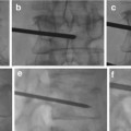
Routine fluoroscopy to accurately locate the center of the femoral head is recommended.
Infiltration of local anesthetic (1–2 % lignocaine).
A subsequent single-wall puncture with a 21-gauge needle is performed with a small skin insertion to aid sheath insertion.
Ultrasound guidance should be readily available and is a valuable adjunct.
Long sheaths 25 cm or more are useful in the setting of diseased or tortuous iliac vessels.
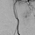
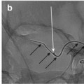
Immediately after sheath insertion a femoral angiogram should be performed. This leads to early discovery of arterial injury or dissection. It also ensures that the puncture site is appropriate for insertion of a closure device.
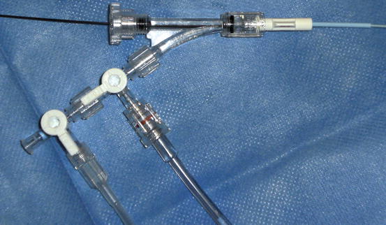
After gaining access to the (right) common femoral artery, the following are routinely used:
Short 4-French sheath
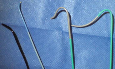
0.035″ hydrophilic angled glide wire
Closed system pressurized saline flush (Fig. 2)