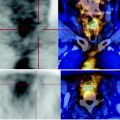
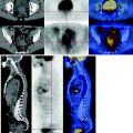
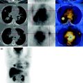
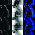
Fig. 27.1
Mass characterized by abnormal glucose consumption at the cervix, SUV max 12. This mass is in contiguity with the anterior wall of the rectum (arrow), infiltrates below the posterior wall of the vagina up to the inferior third and back, in the mesorectal fat
There are not evident adenopathies characterized by pathological metabolism.
No areas of abnormal glucose consumption in the remaining parts of the body examined.
27.4 Conclusions
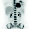
The PET scan shows a mass of the uterine cervix with infiltration of the surrounding tissues with high glucose metabolism (See Fig. 27.2).
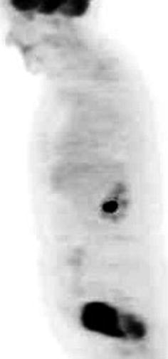
Fig. 27.2


The MIP image shows the intense glucose consumption of the pelvic mass fingerprinting the posterior wall of the bladder
Stay updated, free articles. Join our Telegram channel

Full access? Get Clinical Tree


