Chapter 2
Chest and Cardiovascular Anatomy
 Question 1:
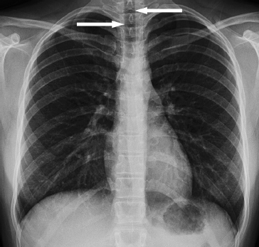
| Name the arrowed structure |
 Question 2:
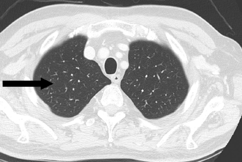
| Name the arrowed structure |
 Question 1: PA chest radiograph
Answer: Trachea
• The trachea is a rigid, tubular structure responsible for the passage of air from the pharynx to the lungs and is reinforced by C-shaped cartilaginous rings. The flat band of muscle and connective tissue posterior to it is called the posterior tracheal membrane, which can appear convex during expiratory views on CT.
• It is 12 to 15 cm long in adults.
• On a PA chest radiograph, it is seen as a midline transradiant cylindrical structure beginning at the level of the lower part of the cricoid cartilage and extending to the carina (from C6 to approximately T5).
• A slight deviation to the right is normal, particularly in children.
 Question 2: Axial CT of the chest
Answer: Right upper lobe
• The right upper lobe is separated from the middle lobe by the horizontal fissure.
• It is divided into three segments: anterior, apical, and posterior.
• The interface between the right upper lobe medial pleural surface and right lateral tracheal border, when seen on a chest radiograph, is termed right paratracheal stripe.
 Question 3:
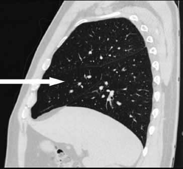
| Name the arrowed structure |
 Question 4:
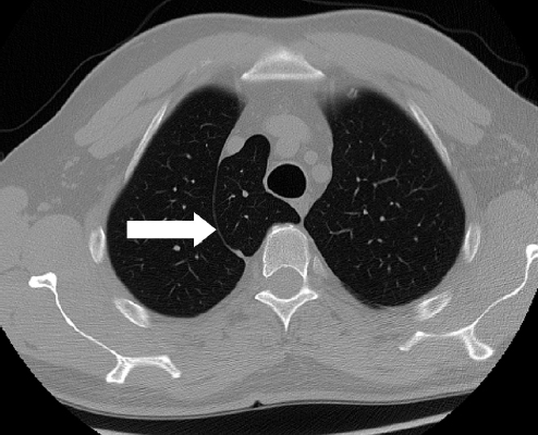
| Name the arrowed structure |
 Question 3: Sagittal CT of the chest
Answer: Middle lobe
• The middle lobe is divided into two segments: medial and lateral.
• It is separated from the upper lobe by the horizontal fissure superiorly and from the lower lobe by the oblique fissure inferiorly.
 Question 4: Axial CT of the chest
Answer: Azygos fissure
• An azygos fissure is the most common accessory fissure and is a common examination question.
• An azygos fissure is seen in approximately 1% of the population.
• It occurs because of failure of normal migration of the azygos vein, which leads to invagination of the parietal and visceral pleura along its course. Sometimes the section of lung medial to the fissure is termed the azygos lobe.
• If complete, it has clinical relevance because it acts as a barrier to apical consolidation extending inferiorly to the rest of the upper lobe.
 Question 5:
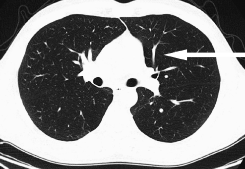
| Name the arrowed structure |
 Question 5: Axial CT of the chest
Answer: Left upper lobe
• The left upper lobe is analogous to a combined right upper and middle lobe.
• It is separated from the left lower lobe by the left oblique fissure.
• It is divided into four segments: anterior, apicoposterior, and the superior and inferior lingular segments.
• The figure below illustrates the different lung segments within the lung lobes with which you should familiarise yourself.
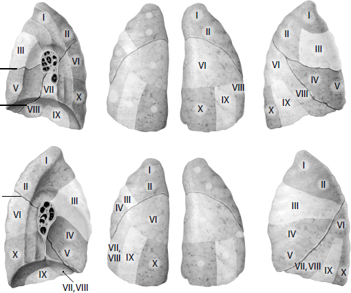
From Atlas of Anatomy, © Thieme 2008, illustrations by Markus Voll.
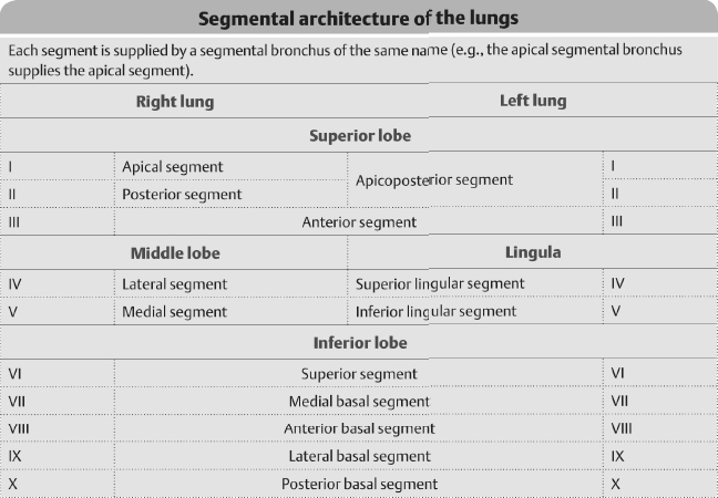
Note: In the left lower lobe, the anterior basal and medial basal segments are conjoined to form the anteromedial basal segment.
 Question 6:
| Name the arrowed structure |
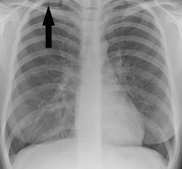
 Question 6: PA chest radiograph
Answer: Companion shadow beside the right clavicle
• The companion shadow is the skin and subcutaneous fat that are seen as a thin soft tissue stripe along the upper edge of the clavicle. It should not be mistaken for an abnormality.
• Similar companion shadows can be formed by the ribs and scapulae. They are not always seen on a radiograph.
 Question 7:
| Name the arrowed structure |
 Question 8:
| Name the arrowed structure |
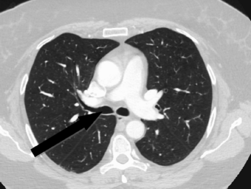
 Question 7: Axial CT of the chest
Answer: Right main bronchus
• The right main bronchus arises from the trachea at the carina.
• It forms a more obtuse angle with the trachea compared to the left (lies at 25° to the median plane).
• It is considerably shorter than the left (about half the length) with a mean length of 2.2 cm.
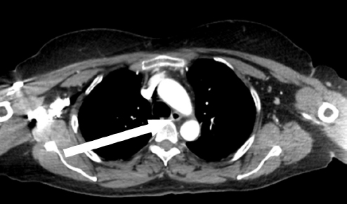
 Question 8: Axial CT of the chest
Answer: Oesophagus
• The oesophagus is the muscular tubular structure that is responsible for conducting food from the mouth to the stomach. It begins at the level of the cricopharyngeus and terminates at the gastro- oesophageal junction and measures approximately 25 cm in length.
• The examination aims to test the candidate on common anatomical structures, but in somewhat unusual modalities or planes such as an axial CT in this instance, or a sagittal CT demonstrating a longitudinal ‘slip’ of air posterior to the trachea that may project in and out of plane.
 Question 9:
| Name the arrowed structure |
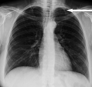
 Question 9: PA chest radiograph
Answer: Left first rib
• The first rib is the flattest and most curved rib.
• The first rib arises from the upward sloping transverse process of the first thoracic vertebra; this distinguishes it from a cervical rib, which arises from the downward sloping transverse process of the seventh cervical vertebra.
 Question 10:
| Name the arrowed structure |
 Question 11:
| Name the arrowed structure |
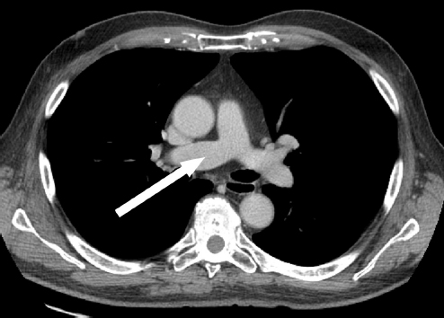
 Question 10: Axial CT of the chest
Answer: Right main pulmonary artery
• The right main pulmonary artery arises at a right angle from the pulmonary trunk and passes behind the ascending aorta and the superior vena cava. It passes anterior to the right main bronchus.
• It is longer than the left main pulmonary artery.
• The left main pulmonary artery arches superiorly over the left bronchus making it much shorter. This is why the left hilar point is higher than the right on a PA chest radiograph.
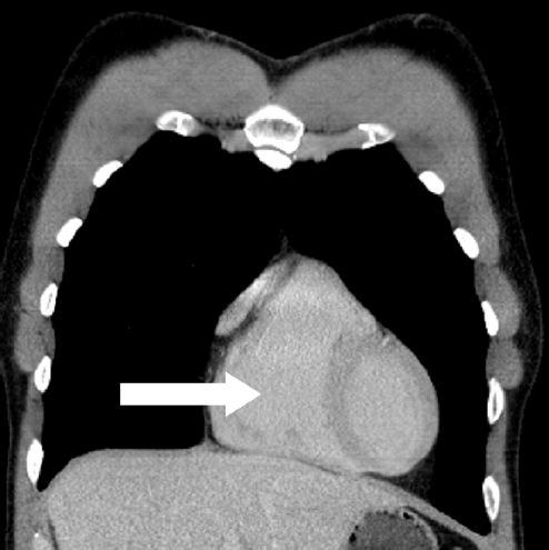
 Question 11: Coronal CT of the chest
Answer: Right ventricle
• The right ventricle is an anterior structure and, when anatomically normal, is not visible on a PA radiograph. It is border-forming on a lateral projection.
• Being the second largest chamber in a normal heart, it is readily identifiable.
• Sometimes the right ventricle will be shown in alternative projections, such as sagittal views where its inferior border usually comes into contact with the sternum, or a coronal view (as in this case) to show its anterior position.
 Question 12:
| Name the arrowed structure |
 Question 13:
| Name the arrowed structure |
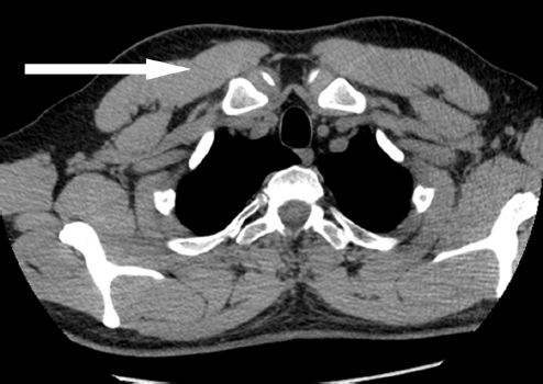
 Question 12: Axial CT of the chest
Answer: Right pectoralis major muscle
• The pectoralis major muscle is a fan-shaped muscle that covers the anterior superior aspect of the thorax and is the most superficial muscle.
• It has two components that are capable of acting independently: a sternal component and a clavicular component. These converge to cross below the shoulder joint to insert at the bicipital groove.
• The pectoralis major muscle is a strong adductor of the arm and also acts as an internal rotator of the humerus.
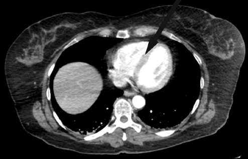
 Question 13: Axial CT of the chest
Answer: Interventricular septum
• The interventricular septum is a strong, thick structure composed of muscular and membranous components that separate the ventricles. It bows to the right due to the higher left ventricular pressure.
• Assessment of the interventricular septum is important because it is a marker of many cardiopulmonary diseases such as pulmonary hypertension, ventricular septal defects, and interventricular aneurysms.
 Question 14:
| Name the arrowed structure |
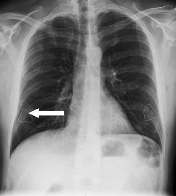
 Question 14: PA chest radiograph
Answer: Right nipple marker
• A nipple marker is a high-density object (usually a metallic triangle or marker) that is placed over the nipple to help differentiate nipple shadows that appear as soft tissue densities from a true parenchymal nodule in erect chest radiographs.
• Nipple shadows, if present, have a characteristic location at the level of the fifth or the sixth anterior rib and often have a well-defined lateral margin; their location can vary with breast size.
 Question 15:
| Name the arrowed structure |
 Question 16:
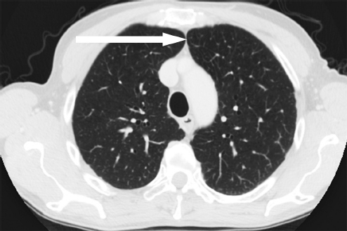
| Name the arrowed structure |
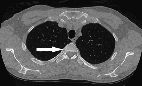
 Question 15: Axial CT of the chest
Answer: Right costovertebral joint
• A costovertebral joint, as the name implies, is the articulation between the head of a rib and the vertebral column.
• A typical rib articulates at two points with the spine: the head of the rib articulates with the vertebral body (costovertebral) and the tubercle of the rib articulates with the transverse process (costotransverse).
• Atypical ribs such as the 11th and 12th ribs do not articulate with the corresponding transverse processes of the vertebral body.
• The costovertebral joint is a synovial joint and, as such, is prone to synovial disease processes such as rheumatoid arthritis.
 Question 16: Axial CT of the chest
Answer: Anterior junctional line
• The anterior junctional line is formed by the visceral and parietal pleura lining the left and right upper lobes.
 Question 17:
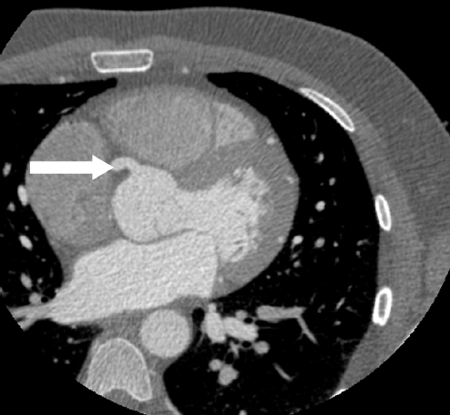
| Name the arrowed structure |
 Question 17: Axial CT of the heart
Answer: Right coronary artery
• The right coronary artery arises from the right coronary sinus (also known as the anterior sinus) and descends in the anterior atrioventricular groove.
Stay updated, free articles. Join our Telegram channel

Full access? Get Clinical Tree


