These are most commonly an osteomyelitis or a neoplastic disease • Malignant rib tumours: these are commonly metastatic deposits or myeloma • Osteomyelitis: this is uncommon • Pleural thickening usually represents an organized end stage of infective or non-infective inflammation • If generalized and gross it is termed a fibrothorax and may cause significant ventilatory impairment • A localized fibrous tumour of the pleura (⅔ are benign)
Chest wall and pleura
CHEST WALL: BONY AND SOFT TISSUE LESIONS
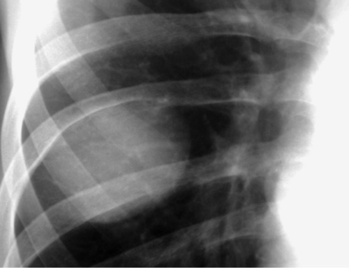
RIB LESIONS
Aggressive
Destructive rib lesions
 primary malignant tumours are rare (but usually a chondrosarcoma)
 it may be due to haematogenous spread (e.g. staphylococcal or tuberculous), or it may be caused by direct spread from the lung or pleural space (e.g. actinomycosis)
DIFFERENTIAL OF RIB NOTCHING
Inferior rib notching
Arterial: Coarctation of the aorta, aortic thrombosis, subclavian obstruction, any cause of pulmonary oligaemia
Venous: Superior vena cava obstruction
Arteriovenous: Pulmonary arteriovenous malformation, chest wall arterial malformation
Neurogenic: Neurofibromatosis (ribbon ribs)
Superior rib notching
Connective tissue diseases: Rheumatoid arthritis, SLE, Sjögren’s, scleroderma
Metabolic: Hyperparathyroidism
Miscellaneous: Neurofibromatosis, restrictive lung disease, poliomyelitis, Marfan’s syndrome, osteogenesis imperfecta, progeria
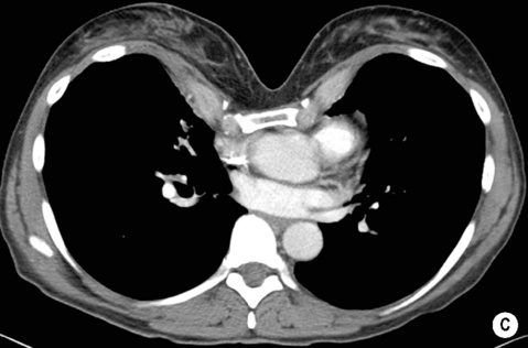
DISEASES OF THE PLEURA
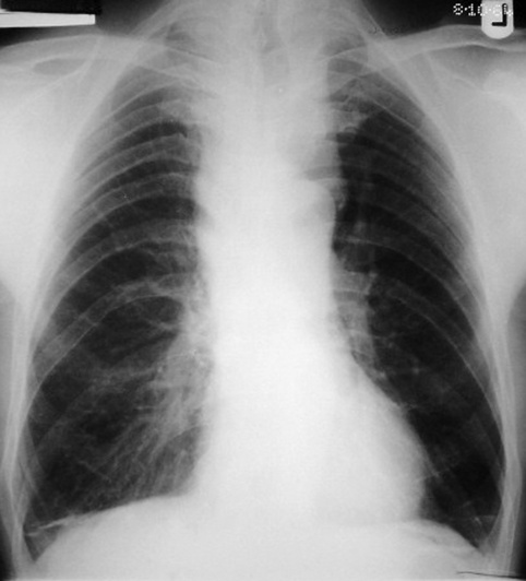
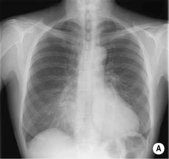
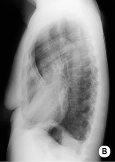
PLEURAL THICKENING AND FIBROTHORAX
DEFINITION
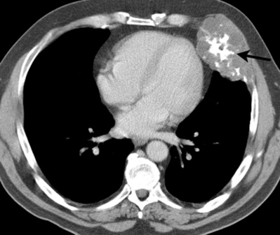
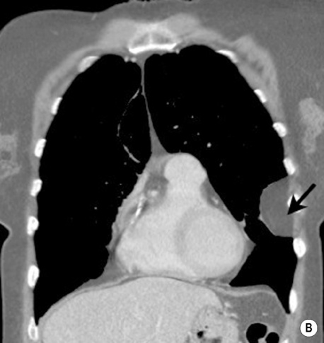
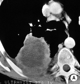
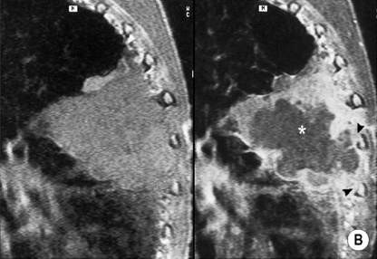
LOCALIZED FIBROUS TUMOUR (LOCALIZED MESOTHELIOMA)
DEFINITION
 there is no relation to previous asbestos exposure
 they are occasionally associated with syndromes (e.g. basal cell naevus syndrome) or other anomalies (e.g. Sprengel’s deformity)
 it can cause a thoracic outlet syndrome and is often bilateral and asymmetrical
 they are most commonly cartilaginous tumours (e.g. a chondroma or osteochondroma)
  they are predominantly found in an anterior location and may show characteristic cartilaginous calcification
 histiocytosis X
  haemangioma
  aneurysmal bone cyst
 MRI can determine the extent of a Pancoast’s tumour (and assess the relationship between the tumour and the plexus brachialis)
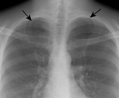
 it is accompanied by ipsilateral hand and arm anomalies (particularly syndactyly)
 soft tissue components suggest a liposarcoma
 T2WI: intermediate SI (and low SI with fat suppression)
 widened intervertebral foramina
 T2WI: high SI
  T1WI + Gad: marked contrast enhancement
 there may be bone remodelling and hypertrophy
 there are signal inhomogeneities generated by vessels, soft tissue and haemorrhage
 T2WI: high SI
 the most common are lipo- or fibrosarcomas
 it may be an isolated abnormality or associated with other disorders such as Marfan’s syndrome or congenital heart disease (particularly an ASD)
 PA CXR: leftward shift of the heart
  an indistinct right heart border simulating middle lobe disease (the sternum replaces aerated lung at the right heart border)
  a steep inferior slope of the anterior ribs
  undue clarity of the lower dorsal spine seen through the heart
 chondrosarcoma
  lymphoma
  metastatic carcinoma
 histiocytosis X
  Paget’s disease
  fibrous dysplasia
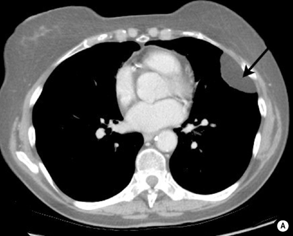

 tuberculosis
  haemorrhagic effusions
 costophrenic angle blunting is common
 US is only reliable if this is > 1cm thick
 pleural thickening is seen particularly on the medial rib aspect
 an asbestos-related fibrothorax is usually bilateral and rarely calcified
 it should be distinguished from a Pancoast’s tumour (if in doubt perform a CT or MRI)
 it can be diffuse but is more often multifocal and often calcified
  it is most commonly found along the lower thorax and diaphragmatic pleura
 they may be calcified