Chest Wall: Scapula


10.1055/b-0034-87922

Chest Wall: Scapula


Table 5.16 Scapula: hypoplasia

Diagnosis


Findings


Comments


Brachial plexus palsy (Erb palsy)


Fig. 5.17a, b


Variable degrees of hypoplasia of the inferior glenoid with posterior subluxation of the humeral head.


Severity may be placed into various categories with cross-sectional imaging.


Recurrent shoulder dislocations


Fig. 5.18


Osseous Bankart and Hill-Sachs lesions.


Recurrent impaction and wear on the anteroinferior glenoid (most commonly) by the humeral head.


Osteonecrosis


Subchondral collapse leads to joint destruction.


DD: sickle cell disease, steroids, radiation and chemotherapy treatments, pancreatitis, etc.


Sprengel deformity


Fig. 5.19a–c, p. 506


Small elevated and rotated scapula ± omocervical (or omovertebral) bone that joins scapula to C5 or C6.


Most common congenital anomaly of the shoulder girdle. Complex anomaly that is associated with malposition and dysplasia of the scapula, regional muscle hypoplasia/atrophy. Associated with Klippel-Feil syndrome.


Dysplasias and syndromes


Achondroplasia, hypochondroplasia, achondrogenesis, campomelic dysplasia, cleidocranial dysplasia, Alpert syndrome.


Glenoid hypoplasia


Hypoplasia of the scapular neck, widened glenohumeral joint space. Bilateral.


± Hypoplasia of other regional structures (humeral head, acromion, clavicle).

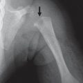
Fig. 5.17a, b Brachial plexus palsy in an 8-year-old. Although the humeral head appears relatively normal, the glenoid is hypoplastic (a) and sloped posteriorly (arrow in b).
Fig. 5.18 Recurrent shoulder dislocations. Osseous Bankart lesion from recurrent shoulder dislocation (arrow) and accompanying intraarticular body in the posterior joint.
Fig. 5.19a–c Sprengel deformity. Klippel-Feil syndrome with small left ribs (a) and a hypoplastic and superiorly located left scapula (Sprengel deformity) that articulates with the neck (b, c).


Table 5.17 Scapula: expansion and destruction

Diagnosis


Findings


Comments


Osteomyelitis


Permeative or moth-eaten pattern of bone destruction.


DD: round blue cell tumors.


Tumors or metastases


Permeative or moth-eaten pattern of bone destruction.


DD: osteosarcoma (new bone formation) and Ewing sarcoma.


Infantile cortical hyperostosis (Caffey disease)


(see Table 5.11 )


Langerhans cell histiocytosis


Lytic lesion or expansile hyperplastic lesion.


May be indistinguishable from osteomyelitis and tumors (round blue cell).

Only gold members can continue reading. Log In or Register to continue

Stay updated, free articles. Join our Telegram channel

Jul 12, 2020 | Posted by in PEDIATRIC IMAGING | Comments Off on Chest Wall: Scapula

Full access? Get Clinical Tree

Get Clinical Tree app for offline access