Osteosclerosis of premature infants and neonates |
Generalized increased density of all the bones. |
Physiologic variant with no clinical signs. Normalizes within the first 3 mo. |
Bone infarcts
Fig. 5.36, p. 519 |
Patchy regions of increased sclerosis that may coalesce. |
Bone infarcts after steroid therapy for inflammatory conditions or tumor treatment. Sickle cell disease. |
Renal osteodystrophy with secondary hyperparathyroidism
Fig. 5.69, p. 542 |
Diffuse sclerosis with trabecular thickening. |
|
Vitamin D–resistant rickets on vitamin D therapy
Fig. 5.62, p. 537 |
Increased density of diaphyses and metaphyses. |
During therapy, sclerosis develops at sites of prior lucency. |
Intrauterine infections |
Increased diaphyseal and metaphyseal density. |
Celery stalk pattern of the metaphyses. Congenital rubella and cytomegalovirus infections. |
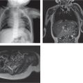
Osteopetrosis (Albers-Schönberg disease)
Fig. 5.67, p. 540
Fig. 5.80, p. 549 |
Generalized increase in bone density. |
Sclerosis obliterates bone marrow preventing hematopoiesis. |
Hypervitaminosis D |
Increased metaphyseal cortical density, particularly at the diaphyseal junction. |
After long-term ingestion of high doses of vitamin D. |
Williams-Beuren syndrome (Williams syndrome) |
(see Table 5.45 ) |
|
Fluorosis |
Combined picture of osteomalacia, osteoporosis, and osteosclerosis. |
Excessive consumption of fluoride. Bone pain and arthralgias. Calcification of ligaments. |
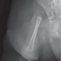
Melorheostosis
Fig. 5.9, p. 498 |
Marked cortical thickening and characteristic dripping candle wax appearance. |
Localized painful swelling and growth disturbances. Follows distribution of dermatomes. |
Primary hypertrophic osteoarthropathy |
(see Table 5.56 ) |
|
Endosteal hyperostosis (van Buchem and Worth types) |
(see Table 5.56 ) |
|
Pycnodysostosis |
Generalized bone sclerosis and mild modeling deformity of the bones. |
Short-limbed dwarfism with generalized increase in bone density. Brittle bones. Acroosteolysis. |
Diaphyseal dysplasia (Camurati-Engelmann disease) |
(see Table 5.56 ) |
|
Erdheim-Chester disease (polyostotic sclerosing histiocytosis) |
|
Usually affects adults. Progressive and widespread patchy sclerosis of the intramedullary region of bones with loss of the corticomedullary junction. Coarse trabecular architecture. Focal rib lesions. |
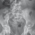
Primary hyperoxaluria (oxalosis) |
Osteoporosis in the early phase and diffuse bone sclerosis in the advanced stage. Subchondral sclerosis in the long bones. Metaphyseal sclerosis, dense epiphyses. |
Growing ends of bones show bulbous enlargement. Pathologic fractures are common. Growth disturbance and increased incidence of urinary calculi due to hyperoxaluria. |