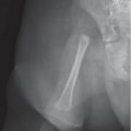
Osteopenia from disuse |
Regional decreased bone density involving immobilized limbs. Cortex is diffusely thinned and the trabeculae are rarified. Metaphyseal ends may be sharply defined. |
Commonly observed during immobilization after trauma/fracture. May accompany soft-tissue atrophy in chronic disuse, arthrogryposis, and other degenerative disorders of muscle and nerves. |
Rickets
Fig. 5.177, p. 618
Fig. 5.178, p. 618
Fig. 5.4, p. 496
Fig. 5.62, p. 537
Fig. 5.69, p. 542 |
Skeletal changes depend on the patient’s age. Wide and hazy growth plates, ill-defined architecture of the trabeculae. Hazy metaphyseal end plates with cupping. |
All forms of rickets (vitamin D–deficiency, renal osteodystrophy, vitamin D–resistance, etc). Cortical erosions occur in secondary hyperparathyroidism. |
Steroid therapy
Fig. 5.113, p. 573 |
Systemic decrease in bone density. Cortex is diffusely thinned and the trabeculae are rarified. |
Decrease in osteoblast activity relative to osteoclasts. |
Hematologic disorders |
Systemic decrease in bone density may be accompanied by changes characteristic for a particular disorder (e.g., hair-on-end skull in thalassemia major). |
Proliferation of cells or deposition of metabolites replaces bone. DD: sickle cell disease, thalassemia major, lysosomal storage disease, leukemia, mastocytosis, mucolipidosis. |
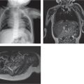
Diffuse metastatic disease
Fig. 5.179, p. 618 |
Diffuse bone demineralization may mask lytic lesions. |
Pathologic fractures. |
JIA |
Periarticular bone demineralization. |
Demineralization may be accompanied by soft-tissue swelling (synovial hypertrophy), joint space narrowing, and/or periarticular erosions. |
Scurvy |
(see Table 5.43 ) |
|
Hyperphosphatasia |
Meshed radiolucent bone texture. |
Progressive skeletal deformity. Enlargement and thickening of the skull and bowed limbs. High levels of serum alkaline phosphatase. |
Congenital hyperparathyroidism |
Pronounced bone demineralization, cortical erosions, and Trümmerfeld fractures in the metaphyses. |
Coarse bone trabeculae, subperiosteal bone resorption, and metaphyseal cupping. |
Osteogenesis imperfecta types I and IV |
Diffuse osteopenia |
|
Idiopathic juvenile osteoporosis |
Disseminated osteoporosis and vertebral body collapse. Presents with bone pain between 8–14 y of age. May spontaneously resolve. |
Homocystinuria |
(see Table 5.45 ) |
|
Metaphyseal chondrodysplasia, Jansen type |
Osteopenia in 70%. Extensive irregularity in mineralization of markedly expanded and cup-shaped metaphyses. |
Relatively preserved epiphyses. The deformity of the metaphyses decreases in adult life. |