The skeleton proximal to the deformity is characteristically normal.
The most common cause of congenital amputation. May be the following etiologies: (1) a true amniotic band constricting the limb and (2) focal and circumferential apoptosis of the soft tissues.
Proximal phocomelia combined with incomplete formation of the distal skeletal segment.
Variable spectrum of limb anomalies arising from maternal ingestion of a drug or toxin that disrupts early fetal skeletal development. Drugs include thalidomide and retinoic acid.
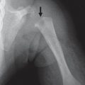
Fig. 5.85a, b Amniotic band syndrome. (a) Amputation of the fingers. (b) A second child with soft-tissue swelling from a constricting band about the tibia (arrow).
Short humerus, dysplastic distally; the femora may also be shortened with resultant rhizomelic dwarfism.
Two forms (isolated to upper limbs or upper and lower with severe dwarfism).
Table 5.50 Dysmelia: radial hypoplasia and aplasia
Diagnosis
Comments
VATER/VACTERL association
The nonrandom association of v ertebral defects, a nal atresia, c ardiac malformations, t racheoesophageal fistula with e sophageal atresia, r adial or r enal dysplasia, and l imb anomalies.
Trisomies 13 (Patan syndrome) and 18 (Edward syndrome)
Characteristic facies, maxillary prognathism, long philtrum, (“carp” mouth), prenatal and postnatal growth retardation, mental retardation, ± upper limb anomalies. Marked variability in presentation.
Poland syndrome
Unilateral absence of hypoplasia of the pectoralis muscle (usually sternocostal portion of the muscle) and a variable degree of ipsilateral hand and digit anomalies.
Congenital heart disease, hand anomalies (triphalangeal thumbs, os centrale), radioulnar synostosis.
Mesomelic dysplasia
Several associated anomalies exist. Langer type is considered to be the homozygote form of the Leri-Weill dyschondrosteosis.
Léri-Weill dyschondrosteosis
Mesomelic dwarfism.
Fanconi anemia, thrombocytopenia-absent radius
Autosomal recessive disorder affecting all bone marrow elements (decrease in one or several hematopoietic cell lines) and associated with cardiac, renal, and limb malformations, and changes in dermal pigmentation.
Fig. 5.86 Cardiomelic syndrome (Holt-Oram syndrome). Absent radius in a patient with Holt-Oram syndrome.
Table 5.51 Dysmelia: ulnar aplasia and hypoplasia
Diagnosis
Findings
Mesomelic skeletal dysplasias
Heterogeneous group of bone dysplasias with disproportionate shortening of the middle segments of the limbs (ulna/radius and/or fibula/tibia). DD: Langer, Nievergelt, Ellis-van Creveld, Maroteaux, Campailla-Martinelli, Reinhart-Pfeiffer, and Robinow syndromes.
Ulnar hypoplasia
May be isolated or associated with other deficiencies (brachydactyly, lobster-claw deformity of feet, mental retardation).
Cornelia de Lange syndrome
Micromelia, phocomelia, hemimelia12 (see Table 5.50).
Nievergelt mesomelic dysplasia
Hypoplasia of the radius and ulna as well as the tibia and fibula. Radioulnar synostosis and a typical rhomboid shape of the tibia and fibula. Mesomelic type dwarfism.
The proximal femur is partially absent, and the entire limb is overall shortened.
The proximal deficiency is a misnomer, as PFFD is often accompanied by aplasia or hypoplasia of the fibula and/or knee (e.g., absence of one or both cruciate ligaments).
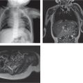
Fig. 5.87 Congenitally short femur.Fig. 5.88a–c Proximal focal femoral deficiency. (a) Radiograph shows the short and malformed proximal femur. T1-weighted (b) and STIR (c) imaging show the relative amount of cartilage in the proximal femur.Fig. 5.89 Proximal focal femoral deficiency with fibular aplasia.
Table 5.53 Dysmelia: tibial aplasia and hypoplasia
Tibial aplasia associated with hand and foot anomalies
Tibial aplasia is associated with clefts, polydactyly, triphalangeal thumbs, and congenital deafness.
Nievergelt types of mesomelic dysplasia
Hypoplastic tibia. Radioulnar synostosis and a typical rhomboid shape of the tibia and fibula.
Werner mesomelic dysplasia
Bilateral hypoplasia of the tibia with polydactyly in the feet and hands.
Table 5.54 Dysmelia: fibular hypoplasia and aplasia
Diagnosis
Findings
Comments
VATER/VACTERL association
The nonrandom association of v ertebral defects, a nal atresia, c ardiac malformations, t racheoesophageal fistula with e sophageal atresia, r adial or r enal dysplasia, and l imb anomalies.