Pelvis: Ischium and Pubic Bones


10.1055/b-0034-87925

Pelvis: Ischium and Pubic Bones


Table 5.21 Pelvis: widening (diastasis) of the pubic symphysis

Diagnosis


Findings


Comments


Trauma


Separation of the medial ends of the pubic bones.


A second pelvic fracture is usually present to complete a break through the pelvic ring.


Osteitis pubis


Lysis of the pubic symphysis and marginal sclerosis during healing phase. Bone marrow edema in the pubis about the pubic symphysis. Increased uptake on bone scan.


Typically occurs in athletes.


Bladder exstrophy


Fig. 5.24, p. 512


Tilted iliac bone with horizontal acetabular roof. True widening of the symphysis.


DD: variants of exstrophy (cloacal, superior vesicle fissure, duplicated exstrophy), epispadias, anorectal anomalies, and urogenital anomalies.


Metabolic


Fig. 5.25, p. 512


Fig. 5.26, p. 512


Margins may appear eroded.


Hyperparathyroidism/renal osteodystrophy.


Infection/inflammation


Lysis and erosions. Widening may be asymmetric.


DD: bacterial, tuberculosis, JIA, reactive arthritis.


Connective tissue disorders


Marfan and Ehlers-Danlos syndromes, for example.


Campomelic dysplasia


Fig. 5.14, p. 501


(see Table 5.12 )


Prune belly syndrome (Eagle-Barrett syndrome)


Flared iliac wings and wide interpubic distance.


Spectrum of congenital anomalies of the gastrointestinal and genitourinary systems. Associated with other syndromes.


Cleidocranial dysplasia


Fig. 5.15a-d, p. 501


Delayed ossification of the pubis.


(see Table 5.15 )


Spondyloepiphyseal dysplasia


Fig. 5.27, p. 512


Absent ossification of the pubis during infancy.


Bone dysplasia with premature osteoarthritis (OA) of the hips. Platyspondyly, short stature. No ossification of the epiphyses of the knees, talus, and calcaneus. Ovoid pear-shaped vertebra in infancy progressing to platyspondyly.

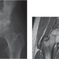
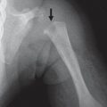
Fig. 5.24 Bladder exstrophy. A 5-month-old with diastasis of the pubic bones from bladder exstrophy.
Fig. 5.25 Metabolic. Widening of the pubic symphysis with lysis in a patient with renal osteodystrophy. Note the erosions in the ischia.
Fig. 5.26 Metabolic. Widened sacroiliac joints and pubic synchondrosis from renal osteodystrophy (arrows).
Fig. 5.27 Spondylometaphyseal dysplasia with changes at the hips and iliac bones and widening of the inferior portion of the pubic symphysis.


Table 5.22 Pelvis: flat and rotated iliac bone

Diagnosis


Findings


Comments


Trisomy 21


Fig. 5.28


Flattened, broad pelvis with flared iliac wings, and decreased acetabular and iliac angles.


An enlarged iliac angle may be seen by fetal US in the second trimester of fetal life.


Exstrophy-epispadias complex


Wide pubic symphysis.


Spectrum of anomalies of the lower abdominal wall, bladder, anterior bony pelvis, and e xternal genitalia.


Sacral agenesis and hypoplasia


Associated abnormalities include deformities of the lower extremities and anomalies of the genitourinary tract, lower gastrointestinal tract, and spine.

Fig. 5.28 Trisomy 21 with flattened broad pelvis (flared iliac wings), six non–rib-bearing lumbar vertebral bodies (11 pairs of ribs not shown), and double bubble of duodenal atresia.


Table 5.23 Pelvis: narrow sacrosciatic notch

Diagnosis


Comments


Achondroplasia


(see Table 5.20 )


Pseudoachondroplasia


(see Table 5.20 )


Thanatophoric dysplasia and variants


(see Table 5.20 )


Short rib-polydactyly syndrome, type I (Saldino-Noonan type)


(see Table 5.20 )


Chondroectodermal dysplasia (Ellis-van Creveld syndrome)


Short limbs, short ribs, postaxial polydactyly, and dysplastic nails and teeth. Sixty percent have congenital heart disease.


Spondyloepimetaphyseal dysplasia


Fig. 5.29a,b


A narrow sacrosciatic notch may be seen in almost all the bone dysplasias with a small iliac bone (see Fig. 5.23 ).

Fig. 5.29a, b Spondyloepimetaphyseal dysplasia at the hips (a) and knee (b).

Only gold members can continue reading. Log In or Register to continue

Stay updated, free articles. Join our Telegram channel

Jul 12, 2020 | Posted by in PEDIATRIC IMAGING | Comments Off on Pelvis: Ischium and Pubic Bones

Full access? Get Clinical Tree

Get Clinical Tree app for offline access