Intermittent progressive heterotopic bone replaces skeletal muscle and connective tissues.
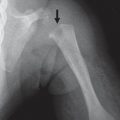
Fig. 5.38 Radioulnar synostosis.Fig. 5.39a, b Isolated ankylosis. (a) Radiohumeral synostosis. (b) US shows a synchondrosis (arrow) where the joint should be located.H HumerusR Radius
Cartilage loss, marginal osteophytes, subchondral bone marrow edema, sclerosis, and cysts.
Although typically seen in adults, secondary OA may complicate JIA or the later stages of AVN. Any disorder that causes abnormal m echanics may lead to OA: posttraumatic, after repair of anterior cruciate ligament.
Joint space narrowing at sites of inflammation. May have imaging findings of synovial thickening and hyperemia, bone marrow edema (MRI), and erosions.
Inflammation contributes to acute cartilage loss.
Hemophilia
Resembles JIA. Epiphyses of affected joints are often enlarged.
Narrowing of the joint space usually results from diseases that cause cartilage loss. A narrowed joint space may be a precursor to ankylosis.
Fig. 5.40 Osteoarthritis. Advanced joint degeneration from slipped capital femoral epiphysis of the left hip with joint space narrowing and osteophyte formation limiting abduction (arrow).Fig. 5.41a, b Juvenile idiopathic arthritis with diffuse joint space narrowing at both hips (a) that is greater on the right. (b) Corresponding coronal T1-weighted image.
Table 5.29 Joints: proximal radioulnar synostosis
Diagnosis
Comments
Congenital
Autosomal dominant and sporadic forms.
Associated skeletal anomalies
Developmental dysplasia of the hip, club feet, missing or diminutive thumb, coalescence of carpal bones, symphalangism, and dislocation of radius.
Associated syndromes
Multiple exostoses, acrocephalopolysyndactyly, Holt-Oram, mandibulofacial dysostosis, Nievergelt dysplasia and Apert, Williams, Klinefelter (and other variants of Klinefelter syndrome with extra sex chromosomes), Nievergelt-Pearlman, and fetal alcohol syndromes.
Proximal radioulnar synostosis may arise from a defect in longitudinal segmentation at the seventh week of development and is bilateral in 60% of cases. Surgery is rarely indicated (because bridge regrows) except for severe pronation deformities.
Only gold members can continue reading. Log In or Register to continue