KEY FACTS
Terminology
- •
Spectrum of extrahepatic and intrahepatic bile ducts malformations characterized by fusiform dilatation
Imaging
- •
Todani classification
- ○
Type I: Solitary fusiform or cystic dilatation of common duct
- ○
Type II: Extrahepatic supraduodenal diverticulum
- ○
Type III: Choledochocele
- ○
Type IVa: Both intrahepatic and extrahepatic cysts
- ○
Type IVb: Multiple extrahepatic cysts without intrahepatic cysts
- ○
Type V: Single or multiple intrahepatic cysts (Caroli)
- ○
- •
Ultrasound
- ○
Cystic extrahepatic mass communicating with common hepatic or intrahepatic ducts
- ○
Intrahepatic ductal dilatation due to simultaneous involvement or secondary to stenosis
- ○
Abrupt change of caliber at junction of dilated segment to normal ducts
- ○
- •
MRCP and ERCP: Best depiction of choledochal cysts and anomalous pancreatobiliary junction union
Top Differential Diagnoses
- •
Biliary obstruction of various causes
- •
Pancreatic pseudocyst
- •
Primary sclerosing cholangitis
- •
Recurrent pyogenic cholangitis
Clinical Issues
- •
Usually diagnosed in childhood (80%)
- •
Triad: Recurrent right upper quadrant pain, jaundice, palpable mass
- •
Complications: Biliary calculi, cholangitis, carcinoma
- •
Surgical excision and enterobiliary reconstruction
Diagnostic Checklist
- •
Rule out other conditions that can cause marked biliary dilatation
Scanning Tips
- •
Caroli disease may appear as unusually shaped cystic structures; look for central dot sign (portal vein within dilated duct) and communication with biliary ducts

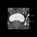
 in a patient with type I choledochal cyst.


