Choledocholithiasis
Kavya E. Reddy
Ellie R. Lee
CLINICAL HISTORY
41-year-old female with right upper-quadrant pain, fever, and vomiting.
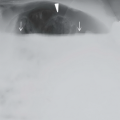
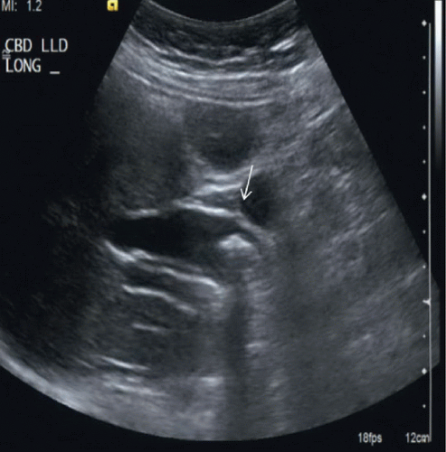 FIGURE 75A |
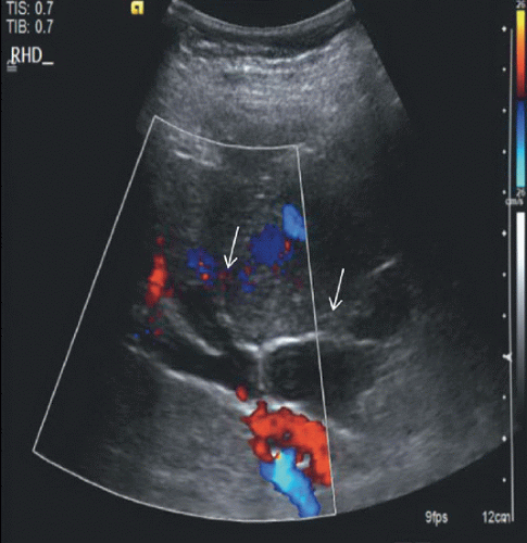 FIGURE 75B |
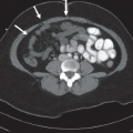
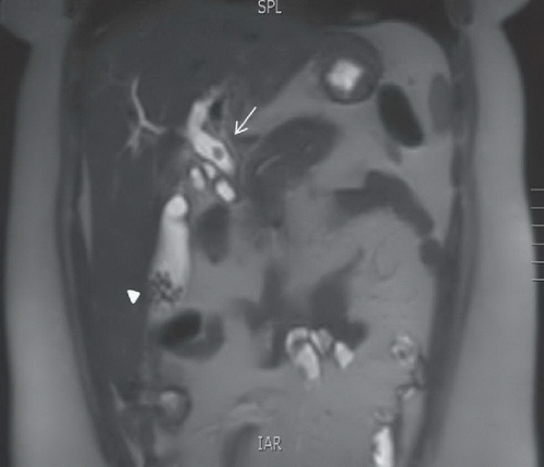 FIGURE 75C |
FINDINGS
Figure 75A: Longitudinal US image of the common bile duct demonstrates an echogenic focus with posterior shadowing in the distal common bile duct (arrow), compatible with choledocholithiasis. The common bile duct is dilated. Figure 75B: Longitudinal color Doppler image of the proximal common bile duct demonstrates dilation of the intra and extrahepatic bile ducts (arrows) proximal to the common bile duct stone. Figure 75C: Coronal T2-weighted MRI image of the abdomen demonstrates a hypointense gallstone in the common bile duct surrounded by hyperintense bile (arrow). Common bile duct dilation is identified. In addition, multiple small hypointense gallstones are identified in the gallbladder (arrowhead).
Related posts:


Stay updated, free articles. Join our Telegram channel

Full access? Get Clinical Tree


