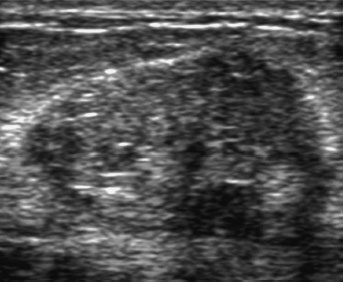
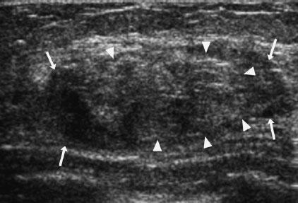
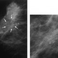
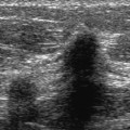
5 Circumscribed Masses: Fat-Containing Masses A 39-year-old woman has just stopped breast-feeding her 10-month-old infant and now finds a new breast lump. • Left breast: 3 cm lump in the upper outer quadrant • Right breast: normal exam Mass (Fig. 5.1) • Margin: circumscribed • Shape: oval • Density: fat-containing Fig. 5.1 In the upper outer quadrant of the left breast, there is a well-defined oval mass. (A) Left mediolateral oblique (MLO) mammogram. (B) Left craniocaudal (CC) mammogram. Frequency • 10 MHz Mass • Margin: well defined • Echogenicity: isoechoic • Retrotumoral acoustic appearance: single edge shadowing • Shape: ellipsoid (Fig. 5.2) Fig. 5.2 Left radial breast sonogram. The mammographic mass identified in Fig. 5.1 corresponds to a well-defined mass with heterogeneous echogenicity. Milky fluid was aspirated from this mass. • Galactocele • Breast Imaging Reporting and Data System (BI-RADS) assessment category 2, benign finding • Galactoceles are benign milk-filled cysts. This lesion is generally discovered either during or shortly after lactation. Mammographically, galactoceles are well-defined and oval and may appear to be completely fat density, heterogeneous density, or equal density to glandular parenchyma. • Sonographically, galactoceles are hypoechoic, isoechoic, or heterogeneous in echogenicity. Fluid debris levels may be present. Jackson VP, Jahan R, Fu YS. Benign breast lesions. In: Bassett LW, Jackson VP, Jahan R, Fu YS, Gold RH, eds. Diagnosis of Diseases of the Breast. Philadelphia: WB Saunders; 1997:357–443 Salvador R, Salvador M, Jimenez JA, Martinez M, Casas L. Galactocele of the breast: radiologic and ultrasonographic findings. Br J Radiol 1990;63:140–142 A 39-year-old woman presents with a new left breast lump. Left breast sonography is initially performed. As a result of the sonogram, a mammogram has been done. • Left breast: 5 cm palpable lump in the upper inner quadrant • Right breast: normal exam Mass (Fig. 5.3) • Margin: circumscribed • Shape: oval • Density: fat-containing Fig. 5.3 At the 9 o’clock position of the left breast, there is a well-defined, oval, fat-containing mass with heterogeneous density (arrows). (A) Left MLO mammogram. (B) Left CC mammogram. Frequency • 10 MHz • Margin: well defined • Echogenicity: heterogeneous • Retrotumoral acoustic appearance: no shadowing • Shape: ellipsoid Fig. 5.4 Left radial breast sonogram. The palpable lump corresponds to a well-defined, oval, solid mass of heterogeneous echogenicity. The peripheral portion of this mass is isoechoic to fat (arrows), and the central portion is hyperechoic (arrowheads). This mass corresponds to the lesion identified in Fig. 5.3. • Hamartoma • BI-RADS assessment category 2, benign finding • Hamartoma is a benign tumor that consists of mature tissues normally present in the breast. However, usually one element dominates the mass. Mammographically, the mass is well defined, but the density of the mass varies with its composition. If the mass does not have much fat, then it may be completely radiopaque. However, if the mass is a mixture of fat and other soft tissues, then it will be mixed density, as in this case. If the mass is completely radiopaque, then the differential diagnosis is fibroadenoma, cyst, or carcinoma. If the mass has mixed density, then the mammogram is diagnostic of hamartoma. • Sonographically, hamartoma has a variety of appearances. The individual components of the tumor should sono-graphically match their corresponding normal elements within the breast. The fatty component is isoechoic to fat, and the fibroglandular elements are hyperechoic to fat. Adler DD, Jeffries DO, Helvie MA. Sonographic features of breast hamartomas. J Ultrasound Med 1990;9:85–90 Anderson I, Hildell J, Linell F, et al. Mammary hamartomas. Acta Radiol 1979;20:712–720 Beatty SM, Orel SG, McCarthy DM, et al. Breast hamartomas: MR appearance. Breast Dis 1995;8:275–281 Cooper RA, Johnson MS, Laissue J-A. Juvenile hypertrophy presenting as a discrete breast mass. Can Assoc Radiol J 1992;43:218–220 Crothers JG, Butler NF, Fortt RW, Gravelle IH. Fibroadenolipoma of the breast. Br J Radiol 1985;58:191–202 Evers K, Yeh I-T, Troupin RH, et al. Mammary hamartomas: the importance of radiologic-pathologic correlation. Breast Dis 1992;5:35–43
 Case 5.1 Galactocele
Case History
Physical Examination
Mammogram

Ultrasound
Pathology
Management
Pearls and Pitfalls
Suggested Reading
 Case 5.2 Hamartoma
Case History
Physical Examination
Mammogram

Ultrasound
Pathology
Management
Pearls and Pitfalls
Suggested Reading
Related posts:


Stay updated, free articles. Join our Telegram channel

Full access? Get Clinical Tree


