| SKULL BASE REGION | Prepontine cistern/trigeminal nerve |
| HISTOPATHOLOGY | N/A |
| PRIOR SURGICAL RESECTION | No |
| PERTINENT LABORATORY FINDINGS | N/A |
Case description
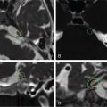
This is a 59-year-old patient with an incidental lesion compatible with a right cisternal trigeminal schwannoma (TS) on brain magnetic resonance imaging (MRI) ( Figure 7.34.1 ). Initial “wait-and-scan” strategy was pursued. During serial follow-up MRIs, the TS exhibited volumetric growth of approximately 30%, without any new symptoms. Gamma Knife radiosurgery (GKR) treatment was then performed ( Figure 7.34.2 ).
| Radiosurgery Machine | Gamma Knife – Icon |
| Radiosurgery Dose (Gy) | 12, at the 55% isodose line |
| Biologically effective dose (Gy) | 72.97 Gy |
| Number of Fractions | 1 |

From left to right: T1-gadolinium injected MRI in coronal, axial, and sagittal planes showing a lesion centered at the cisternal part of the trigeminal nerve, without contact with the brainstem.

From left to right: T1 noninjected MRI, T1-gadolinium injected MRI, bone CT in the axial plane. The dosimetry is colored in yellow and corresponds to the 12-Gy dose prescription. We always use multimodal imaging for target definition, which may also include T2 CISS/Fiesta sequences.
| Critical Structure | Dose Tolerance |
|---|---|
| Brainstem | Marginal dose of 12 Gy due to absence of direct contact between TS and brainstem; the risk of adverse radiation events (ARE) at the brainstem level is virtually zero |
Related posts:
 Esthesioneuroblastoma – delayed postoperative radiosurgery for recurrence at long-term
 Null cell – delayed postoperative radiosurgery for growing perioptic residual
 Chondrosarcoma – definitive radiosurgery after subtotal resections
 Trigeminal neuralgia due to microvascular conflict – upfront radiosurgery
 Capillary hemangioma – postoperative radiosurgery for residual tumor
 Superior sagittal sinus meningioma – delayed postoperative, multisession radiosurgery for growing residual
Stay updated, free articles. Join our Telegram channel

Full access? Get Clinical Tree


