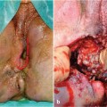
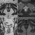
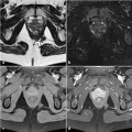
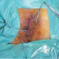
Fig. 9.1
Anamnesis room
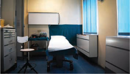
Fig. 9.2
Physical examination room
A complete physical examination must always be performed. Abdominal tenderness, hepatomegaly, splenomegaly, groin adenopathy, should be identified and described. In a patient with Crohn’s disease, abdominal masses can be a sign of ileal dilatation, intra-abdominal abscess, or mesenteric adenopathy. In such patients, enterocutaneous fistula or multiple abdominal scars can be observed. Any doubt during the clinical exam should be pursued with the proper imaging test.
The perineal exam is based on three steps:
Inspection and palpation of the perianal skin
Digital exploration
Examination of muscular function
The patient can be placed in different positions so as to facilitate the examination:
Sis’m position: The patient lies on his or her left side with the buttocks near the edge of the examination table and knees in flexion (Fig. 9.3).9 Clinical Examination
Lithotomy position: The patient lies supine, with the perineum positioned at the edge of the examination table and the feet above or at the same level as the hips.
Stay updated, free articles. Join our Telegram channel

Full access? Get Clinical Tree


