Chapter 4 Clinical Methods
Ultrasound Biomicroscopy
Introduction
Ultrasound biomicrosopy (UBM) utilizes high frequency ultrasound to evaluate anterior ocular structures (Chapter 2).1–5 As in the examination of the posterior segment with B-scan and A-scan, the specific type of examination performed is determined by the indication for examination. UBM has numerous clinical applications, particularly in the evaluation of corneal diseases (Chapter 8), glaucoma (Chapter 9), anterior segment tumors (Chapter 11), and trauma (Chapter 16). Modified UBM is also increasingly used for measurements of organs in laboratory animals (Chapter 17). Digital UBM is an important tool in assessment of patients undergoing refractive surgical procedures (Chapter 6). This chapter describes the methods for performing ultrasound biomicroscopy.
Basic positioning and patient preparation
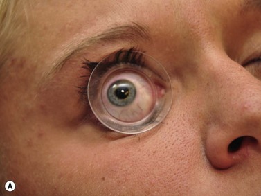
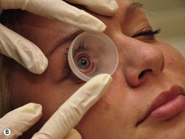
Positioning of the patient for UBM examination is similar to that of contact B-scan and diagnostic A-scan in the evaluation of the posterior segment. It is recommended to recline the patient to the supine position and place an anesthetic drop in the eye to be examined. A fluid immersion technique is necessary to provide the stand-off distance required to view the anterior ocular structures. Specially designed immersion UBM scleral shells are most commonly used to hold the eyelids open and provide a reservoir for the coupling agent (Figure 4.1).1 Water is most easily used in the shells as a coupling agent; however, fluid loss and air bubble obstruction during the procedure can hinder the efficiency of the examination. Methylcellulose is a good alternative because it has low sound attenuation and greater viscosity, preventing fluid loss during the examination.
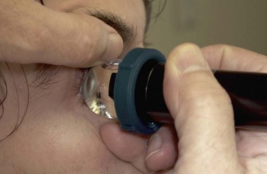
The UBM probe does not have a membrane over the probe tip covering the moving transducer (Chapter 2). Such a membrane would cause significant sound attenuation at the high frequencies used. Therefore, the examiner must be extremely careful to avoid contact with the cornea. Recently, a single use ultrasound cover, ClearScan® has been developed.6 It consists of an extremely thin acoustically invisible film that can be filled with water and placed over the probe tip of the UBM (Figure 4.2). It protects the cornea from the moving transducer, and allows for a thorough examination without the discomfort and peripheral examination constraints of a scleral shell.
UBM probe positions
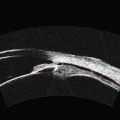
A typical, 50 mHz UBM probe has a penetration depth of approximately 5.0 mm and a resolution of 37 µm. The relationship of the probe position and display image for the UBM is different than that of the conventional contact B-scan. During UBM examination, the probe is placed directly over the anterior ocular structures to be imaged. Structures that are closest to the probe tip are at the top of the screen, those farthest away are at the bottom (Figure 4.3
Stay updated, free articles. Join our Telegram channel

Full access? Get Clinical Tree


