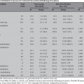
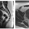
22 Clinical Perspective: Spine Interventions (Orthopedic Surgery) Burt Yaszay and Jeffrey M. Spivak Every year in the United States, more than 700,000 vertebral compression fractures occur secondary to osteoporosis.1 This is in addition to those that are attributed to metastatic vertebral lesions. Significant morbidity can result from both the related pain and deformity associated with these compression fractures. Studies have shown that vertebral compression fractures can lead to decreased physical function, quality of life, and even survival.2,3 The majority of patients that suffer a compression fracture experience acute pain at the time of initial injury. This pain may be initially severe; however, it typically resolves in most patients over the course of weeks to months. Many patients respond to mild analgesics and remain quite functional during this acute episode. However, for some patients the pain can be incapacitating in nature and can ultimately become a chronic problem. Although not completely understood, the development of chronic pain in association with a vertebral compression fracture can be multifactorial.4 There may be progression of the fracture with further bony collapse. Incomplete healing of the fracture can lead to a pseudoarthrosis and a sense of instability or motion through the vertebrae. Finally, the kyphotic deformity that often results from a compression fracture can alter the biomechanics of the spine leading to fatigue of spinal extensor muscles and strain on other dynamic stabilizers of the axial skeleton. Severe kyphosis can also result in discomfort from impingement of the rib cage on the pelvis. In addition to pain, the kyphotic deformity resulting from a compression fracture can have other negative clinical consequences for the patient. Kyphosis shifts the sagittal vertical axis (patient’s center of gravity) anteriorly. This loss of sagittal balance and a decrease in vertebral height can cause a loss of physical height, diminished pulmonary function, compression of abdominal organs, and poor appetite.2 This is supported by the increased mortality rate from pulmonary complications that is seen in elderly women with five or more thoracic compression fractures.5 In addition, kyphosis displaces compressive forces anteriorly on other vertebral bodies, which can lead to additional fractures. The risk of developing a new fracture is increased following a vertebral compression fracture, especially at adjacent levels. Psychosocial effects include limitations in functional abilities and a poor perception of general health.2 Because the symptoms resolve in the majority of patients with a compression fracture, the mainstay of treatment is nonoperative and includes analgesics, bracing, and progressive mobilization.6 Medical evaluation and management of the patient’s osteoporosis must be undertaken if not done previously, to help minimize the risk of additional fractures. For those patients that have failed nonoperative management due to severe incapacitating pain, progressive collapse and/or wedging, or chronic pain with nonunion, more invasive procedures of vertebral cement augmentation can be very helpful to provide the desired outcome of pain relief. Some physicians caring for patients with osteoporotic vertebral compression fractures prefer to be even more aggressive, recommending cement augmentation early in the course of all fractures, to not miss those fractures which will go on to progressive collapse and wedging and become too severe for such treatment. Since the 1990s, percutaneous vertebral augmentation has become increasingly popular for the treatment of these refractory cases.7 Originally described for the treatment of malignant vertebral lesions, vertebroplasty refers to the percutaneous injection of a hardening substance (a “cement”), which is injected into the structurally compromised vertebral body and then hardens, providing immediate stability and augmenting the strength of the vertebral body.8 As far back as the 1980s, it was noted that patients that had acrylic cement injected to strengthen vertebral bodies with angiomas also had an analgesic effect with improvement in their pain. As a result, the indications for vertebroplasty were expanded to include painful osteoporotic vertebral compression fractures. Introduced clinically in 1998, kyphoplasty is similar to vertebroplasty in that it involves the percutaneous injection of acrylic cement into a mechanically compromised or fractured vertebral body.9 However, prior to the injection, a percutaneous inflatable balloon tamp is placed within the vertebral body and inflated, achieving additional restoration of vertebral collapse and wedging and creating a central cavity with impacted bone margins. The subsequent void can then be filled with partially cured (thicker or more viscous) cement through a low pressure injection. This may diminish the risk of unwanted extravasation of cement that is seen more commonly with vertebroplasty. The most commonly used material in vertebroplasty or kyphoplasty is polymethylmethacrylate (PMMA). Typically referred to as acrylic bone cement, it is initially a liquid when first mixed that then polymerizes and hardens through an exothermic reaction. It is felt to be bioinert and does not reabsorb over time. It is also much more rigid (lower modulus of elasticity) than the native vertebral body bone. The indications for vertebroplasty and kyphoplasty continue to evolve. In our practice, the most common clinical use for percutaneous vertebral augmentation is the treatment of painful or kyphotic vertebral compression fractures from osteoporosis or osteolytic lesions. Vertebroplasty is used primarily to relieve pain despite the fact that the mechanism for the pain relief associated with this procedure is not completely understood. One possible explanation is that the bone cement provides immediate immobilization of the fracture and augments the mechanical strength of the affected vertebrae. Another theory is that the heat from the PMMA’s exothermic reaction may desensitize the fractured bone. Although the mechanism may be multifactorial, partial to complete pain relief commonly occurs within 24 to 72 hours.10 Vertebroplasty does not have any significant ability to improve vertebral height that is intrinsic to the procedure. It relies more on the dynamic mobility that is seen with many of these fractures.11 Proper positioning of a patient for the procedure in an antikyphotic position may partially restore vertebral height and improve wedging, especially in relatively acute or unstable fractures. This increase can then be maintained by the solidified bone cement. In addition to providing significant pain relief equal to that of vertebroplasty, kyphoplasty has the added capability of decreasing kyphosis and restoring vertebral body height through the use of a single or paired inflatable balloon tamp.9 Inflation of the properly positioned balloons elevates the endplate and creates a walled-off cavity, which is then supported by the injected cement. If performed within 3 months of the fracture occurrence, kyphoplasty has been shown to diminish kyphosis by up to 50%.12 Therefore, in light of these benefits, we consider kyphoplasty to be the preferred technique for the kyphotic compression fracture. The original indication for percutaneous vertebral augmentation is in the management of painful metastatic vertebral lesions.6 Again, it is unclear how augmentation procedures result in pain control for these patients. Neoplasms are not usually innervated and therefore are not directly painful. Instead, the pressure from the expanding mass or the impending fracture of the weakened bone may be the source of pain. In addition, the tumor cells may secrete local mediators that induce pain. Similar to what is observed in compression fractures, stabilization and desensitization of the bone may be responsible for the diminished pain. An additional source of relief may be from an antitumoral effect of the injected cement. Whether from heat production, toxicity of the PMMA polymers, or local ischemia, a direct cytotoxic effect has been suggested by the decreased local recurrence rate following percutaneous vertebral augmentation.13 Interestingly, the relief of pain following vertebroplasty has not been correlated with the type of lesion or the amount of PMMA injected into the vertebral body.14 Excluding those patients that suffer an associated compression fracture, deformity correction is not typically needed in patients with a metastatic vertebral lesion. Therefore, vertebroplasty has been the treatment of choice in the published literature. Some studies demonstrate sustained pain relief in greater than 70% of the treated cancer patients.15,16
Patient Selection
Indications and Contraindications
![]()
Stay updated, free articles. Join our Telegram channel


Full access? Get Clinical Tree


