Clinical Vascular Examination


Clinical Vascular Examination


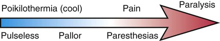
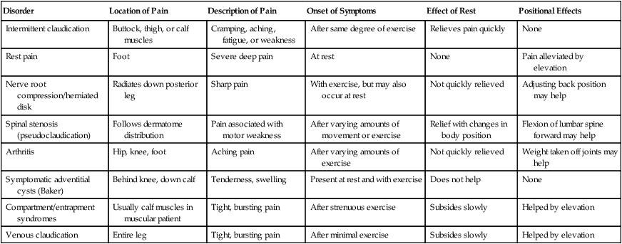
Benjamin S. Brooke and James H. Black, III


Symptoms relating to vascular insufficiency are common complaints among patients presenting to emergency departments in westernized nations. Unfortunately, these symptoms are often misdiagnosed because of variability in presentation and overlap with other diseases. Vascular diseases may present and manifest in a number of different ways, but most can be evaluated after careful and thorough clinical examination. Although atherosclerosis will be asymptomatic in many patients, its natural progression may be identified by a patient’s symptoms, which range from intermittent claudication to ischemic rest pain and gangrene. And although the majority of signs and symptoms result from chronic arterial insufficiency, it becomes essential to recognize acute symptoms that may be limb threatening and require immediate intervention. Moreover, there are many symptoms of nonvascular etiology that mimic vascular insufficiency. It is important to know how to recognize and differentiate symptoms attributed to these other organ systems and to make a differential diagnosis.


The goal of the clinical vascular examination is to identify the cause of the patient’s symptoms while localizing the specific lesion and judging its relative severity. The astute clinician should be able to make a presumptive diagnosis after the examination, formulate a treatment strategy, and prevent the patient from undergoing unnecessary tests that are costly and may have additional risks. Although technology has greatly facilitated the ability to identify and localize vascular disease, it should not be relied on to make the diagnosis. The use of imaging modalities should merely confirm the clinician’s diagnosis after the examination is complete. For some patients, in particular those with acute presentation, the decision to go to the operating room or angiography suite for intervention often has to be made by the end of the examination, without any adjuvant studies.


Detecting vascular disease is a comprehensive process that requires careful review of the medical history to reveal significant symptoms and risk factors, detection of relevant physical examination findings, and use of appropriate supplementary tests to confirm the diagnosis. A systematic head-to-toe approach should be undertaken with every patient, because many individuals will have multiple areas of vascular disease. In this chapter we review the important elements of the clinical vascular examination, including history and physical examination, risk factors, and bedside procedures that can be used to confirm a diagnosis. Patterns of vascular disease are distinguished by anatomic and physiologic causes and differentiated from other conditions. In addition, relevant scientific studies that validate important findings of the history or physical examination are discussed.


History


Obtaining an accurate history of symptoms and their chronology is a critical initial component of a vascular examination. Often a presumptive diagnosis can be made after a detailed history even before the patient has been examined. Or conversely, a patient is referred with an established diagnosis that is determined to be inaccurate after a careful history and examination. Every patient must be queried as to the location of pain, duration of symptoms, whether the symptoms are intermittent or consistent, whether certain circumstances aggravate or alleviate symptoms, and the time relationship to other medical events. Knowing what questions to ask can help discriminate the severity and seriousness of disease as well as guide the diagnostic workup. It is imperative to discriminate symptoms in the history that indicate whether the disease is acute or due to chronic vascular insufficiency. Moreover, knowing a patient’s history and risk factors can help distinguish whether symptoms are caused by an embolus or thrombosis. The presentation and patterns of symptoms for specific types of peripheral vascular diseases are discussed next in more detail.


Chronic Arterial Insufficiency


The natural history of arteriosclerosis in the lower extremity can be viewed as a progression of symptoms that reflect the degree of vessel occlusion and presence of collateral circulation. Symptoms classically begin with an intermittent and reproducible pain termed claudication, a derivation of the Latin word for “limp.” Intermittent claudication is characteristically brought on by a given degree of exercise and relieved after a few minutes of rest. These symptoms reflect a demand for blood flow increases that cannot be met because of atherosclerotic narrowing in one or more arterial segments. Compression of stenotic arteries by exercising muscle and lack of compensatory vasodilation in diseased segments may also contribute to limited extremity perfusion. This pain is often described as aching, cramping, weakness, or fatigue and may present in the buttocks, thighs, calves, or feet. This diagnosis must be considered with any exertional limitation of the lower extremity muscles or any history of walking impairment that gets better shortly after rest. However, there are a number of other causes of extremity pain that must be considered and excluded from the diagnosis (Table 15-1). Among patients with claudication, 25% will experience worsening symptoms, 5% will eventually require revascularization, and only 1% to 2% will come to major amputation.1


TABLE 15-1


Differential Diagnosis of Extremity Pain


Disorder Location of Pain Description of Pain Onset of Symptoms Effect of Rest Positional Effects
Intermittent claudication Buttock, thigh, or calf muscles Cramping, aching, fatigue, or weakness After same degree of exercise Relieves pain quickly None
Rest pain Foot Severe deep pain At rest None Pain alleviated by elevation
Nerve root compression/herniated disk Radiates down posterior leg Sharp pain With exercise, but may also occur at rest Not quickly relieved Adjusting back position may help
Spinal stenosis (pseudoclaudication) Follows dermatome distribution Pain associated with motor weakness After varying amounts of movement or exercise Relief with changes in body position Flexion of lumbar spine forward may help
Arthritis Hip, knee, foot Aching pain After varying amounts of exercise Not quickly relieved Weight taken off joints may help
Symptomatic adventitial cysts (Baker) Behind knee, down calf Tenderness, swelling Present at rest and with exercise Does not help None
Compartment/entrapment syndromes Usually calf muscles in muscular patient Tight, bursting pain After strenuous exercise Subsides slowly Helped by elevation
Venous claudication Entire leg Tight, bursting pain After minimal exercise Subsides slowly Helped by elevation


image


Adapted from Dormandy JA, Rutherford RB. Management of peripheral arterial disease (PAD). TASC Working Group. TransAtlantic Inter-Society Consensus (TASC). J Vasc Surg 2000;31(Suppl):S1–S296.


A query of the specific distribution of claudication reported by the patient is an important focus of the history. Symptoms typically occur in the muscle groups immediately distal to the region of stenosis. The exact site(s) of discomfort or pain should be recorded, along with the relationship of this pain to rest or degree of exertion. Narrowing of the aortoiliac vessels classically presents as the constellation of buttock pain, impotence, and lower extremity muscle atrophy known as Leriche syndrome. Similarly, occlusion of the femoral, popliteal, or proximal tibial arteries will present as exertional calf pain. Multiple studies have found intermittent claudication to be a reliable symptom in the diagnosis of peripheral arterial disease. A recent meta-analysis found that the presence of claudication increased the likelihood of arterial disease being present by over threefold, whereas its absence lowers the likelihood by half that moderate to severe disease is present.2


Rest pain represents progression of ischemic disease in the lower extremity and is characterized by pain in the foot or calf that is made worse by elevation and is generally relieved by placing the extremity in a dependent position to allow gravity to assist in blood flow. This pain may start as a dull aching sensation in the toes that progresses to severe burning pain at night when the patient is lying supine. Patients may describe hanging a foot or limb over the bed or sleeping in a chair to obtain relief. The pain may intensify with cold exposure and be relieved somewhat with application of heat, although strong analgesics are usually needed to manage the discomfort. Skin ulceration and gangrene occur when the minimal nutritional and metabolic requirements of the extremity are not met by adequate blood flow. Usually, multiple levels of arterial disease (iliac, femoral, tibial) are present for this critical ischemia to occur. The pain associated with this process may intensify initially but then diminish or disappear as tissue necrosis involves peripheral sensory nerves.


Acute Arterial Insufficiency


The clinical presentation of acute arterial insufficiency and limb ischemia is characterized by new-onset extremity pain with concurrent changes in neurologic function. Pain is usually the first symptom to be described and is characteristically elicited by passive flexion or stretch of the extremity. This pain is not usually localized exclusively to the distal foot and not affected by limb position, which helps differentiate it from the rest pain of chronic arterial insufficiency. Sensory loss is the earliest neurologic sign; it may be very subtle at first but progresses quickly to frank paresthesias. When extremity perfusion continues to be compromised, muscle strength and motor control become diminished. This spectrum of clinical symptoms and findings are termed the 6 Ps: pain, pallor, poikilothermia, paresthesias, paralysis, and pulselessness (Fig. 15-1).


The onset and pattern of these symptoms is an important clue to the cause of obstruction of blood flow. The diagnosis of arterial embolism is suggested by sudden onset of severe pain in an extremity not previously affected with claudication or other symptoms of obstructive arterial disease. This acute pain may be described as crippling and often may leave the distal extremity weakened. A careful review of the patient’s history will usually reveal the source of emboli or associated risk factors. This includes ventricular thrombi generated weeks to months after a myocardial infarction, or during atrial fibrillation with or without adequate anticoagulation. Other origins of emboli include deep venous thrombus with a patent foramen ovale, mechanical heart valves, endocarditis, or aneurysms located in the aorta or infrainguinal arterial system.


Acute arterial thrombosis is the other main cause of acute insufficiency and is frequently superimposed on an already narrow atherosclerotic lesion. The pain associated with thrombosis is usually not as abrupt, typically following a crescendo course. Classically, a history of claudication or poor circulation is obtained in these patients. Severity and duration of symptoms often correlate with the presence of collateral circulation in the region of the narrowed vessel, helping restore distal perfusion. In the absence of collateral vessels, the pain of thrombosis may be similar to embolic disease. It is important to assess whether the patient has an antecedent coagulation disorder that may predispose to arterial thrombosis. In comparison, patients with deep vein thrombosis may have complaints related to swelling, erythema, and dull pain in the affected extremity. The presence of palpable pulses and/or signs of adequate perfusion in the affected extremity helps differentiate venous thrombosis from an arterial cause.


Cerebrovascular Disease


Patients presenting with carotid disease usually have had a recent transient ischemic attack, stroke in evolution, or completed stroke with persistent neurologic deficits. Transient ischemic attacks may include an account of transient monocular blindness or amaurosis fugax. These brief visual impairments are often described by patients as a “fog,” “shade,” or “curtain over the eye.” When neurologic deficits are fixed, their distribution should be correlated with a point of occlusion in the cerebrovascular circulation. It is important to document the location and chronology of neurologic symptoms in all these patients. A great number of patients with carotid artery disease will be asymptomatic at presentation but may be identified by certain clues in the history. A recent report of nonspecific visual changes may be the first clue that a patient has significant carotid stenosis. Other sources of embolic disease must also be ruled out, including a patient history of cardiac arrhythmias or known patent foramen ovale.


Dec 23, 2015 | Posted by in INTERVENTIONAL RADIOLOGY | Comments Off on Clinical Vascular Examination

Full access? Get Clinical Tree

Get Clinical Tree app for offline access