KEY FACTS
Terminology
- •
Clubfoot (talipes equinovarus)
- ○
Foot is turned in at ankle (varus)
- ○
Foot is pointed (equina)
- ○
- •
Rocker-bottom foot (congenital vertical talus)
- ○
Back of foot is convex (like bottom of rocking chair)
- ○
Toes are upturned
- ○
Imaging
- •
Clubfoot: 2/3 bilateral
- ○
Frontal tibia/fibula view best to show turned-in foot
- ○
Plantar foot view shows short angulated foot
- ○
2/3 are isolated: No other anomalies
- ○
Key associations: Spina bifida, arthrogryposis, aneuploidy, oligohydramnios
- ○
- •
Rocker-bottom foot: 2/3 bilateral
- ○
Sagittal view best to show morphology
- ○
Rarely isolated
- ○
Key associations: Aneuploidy (particularly trisomy 18), spine anomalies, arthrogryposis
- ○
Clinical Issues
- •
Clubfoot is most common musculoskeletal anomaly
- ○
1-3:1,000 newborns
- ○
Prognosis depends on associated anomalies
- ○
Most do not need surgical correction
- ○
- •
Rocker-bottom foot requires more significant intervention
Scanning Tips
- •
3D helps show severity
- ○
Bone-rendered views are additive
- ○
- •
Beware of transient foot position
- ○
Wait for feet to move away from uterus
- ○
Avoid 3rd-trimester diagnosis if possible
- –
Higher false-positive rates
- –
- ○
- •
Look carefully at spine, upper extremities, and fetal movement when lower extremity anomaly diagnosed
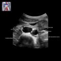
 and a normal left foot
  . 3D images allow for better appreciation of the severity of the clubfoot and allows the patient and orthopedic team to truly see the anomaly.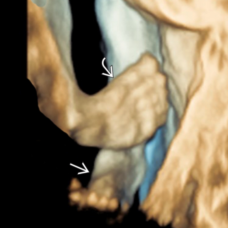
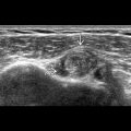
 , classic varus deformity of clubfoot. The finding was isolated and genetic testing was not done. Bilateral isolated clubfoot does not increase risk of aneuploidy over unilateral.
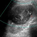
 , which pushes the heel posteriorly
  and bows the sole, creating a rocker-bottom appearance. The correlative ultrasound shows the rocker-bottom heel
  and upturned toes
  .
.Related posts:

Stay updated, free articles. Join our Telegram channel

Full access? Get Clinical Tree


