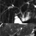
53 Common Incidental Findings Incidental findings within the sinuses are commonly seen on head and neck MRI performed for other reasons. Mucous retention cysts most commonly occur in the maxillary sinuses and are asymptomatic unless they disrupt mucociliary clearance. The typical appearance is illustrated in Figs. 53.1A,B where bilateral lesions exhibit high and low SI on (A) axial T2WI and (B) contrast-enhanced T1WI, respectively. A large lesion can be confused with an air-fluid level, leading to a false diagnosis of sinusitis. The nondependent location of the retention cysts in Figs. 53.1A,B aids in this distinction, but dependent mucous retention cysts are more problematic: a convex border with surrounding air suggests a retention cyst versus the concave up borders of an air fluid level. Evaluation of the lesion in multiple planes can further aid in differentiation. Polyps are also typically asymptomatic and exhibit variable SI based on their relative protein content. Such lesions are not reliably distinguished from mucous retention cysts by their MRI appearance. Mucosal thickening is often asymptomatic, and is defined as thickening of the lining of the maxillary and ethmoid sinuses greater than 4 and 2 mm, respectively. Figures 53.2A and 53.2B demonstrate left maxillary sinus mucosal thickening: compared with the right sinus wall, added moderate and high SI is present on the left in (A) T1WI and (B) T2WI, respectively. Such findings correlate with inflammatory edema or hyperplasia. Abnormal SI within the ethmoid air cells has been shown to alternate from the left to right side over the course of the day as part of a normal nasal cycle. Sinusitis, further discussed in Chapter 56
![]()
Stay updated, free articles. Join our Telegram channel

Full access? Get Clinical Tree


