 The Magnet
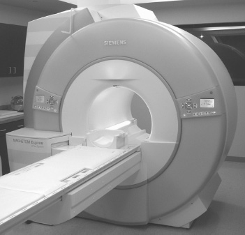
Fig. 1.1
Hydrogen atoms (and of particular relevance, those in water molecules) have a nuclear spin, and associated with the nuclear spin is a magnetic moment. An externally applied magnetic field causes these magnetic moments to preferentially align parallel to the field. The magnetic field strength B0 is measured in tesla (T). A 1.5 T system (Fig. 1.1) provides a magnetic field of ~30,000 times that of the earth, with no permanent effects on human physiology and negligible temporary alterations. The magnetic field is generated by feeding ~2400 A (Fig. 1.2) into the windings of a superconducting magnet. Superconductivity means that once the current is applied, the power supply can be disconnected, the end and beginning of the coil windings connected, and the current will continue to flow. Thus such a magnet will be at field at all times, even during a power outage.
 The Transmitting Radiofrequency Coil
Stay updated, free articles. Join our Telegram channel

Full access? Get Clinical Tree


