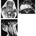
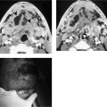
Chapter 175 Congenital anomalies of the third branchial complex are unusual lesions that can either consist of fistulas, sinuses, or cysts. There is no reported gender predilection. Because the third branchial pouch gives rise to a portion of the parathyroid glands and thymus, anomalous embryogenesis may result in a multisystem disease (DiGeorge’s syndrome) rather than an isolated anomaly. The cartilage of the third branchial arch forms the lower body and greater horns of the hyoid bone. The musculature derived from the third branchial arch is limited to the stylopharyngeus muscle, which is supplied by the glossopharyngeal nerve. The mucosa covering the posterior third of the tongue base is also a derivative of the third arch. Some authors claim that the palatopharyngeus muscle and a portion of the skin over the carotid artery are also third arch derivatives. The third branchial cleft is normally obliterated by overgrowth of the second branchial arch. Other derivatives include the inferior parathyroid and thymus glands. The course of a third branchial apparatus fistula can be predicted based on the embryogenesis of the branchial arches and is illustrated in Figure 175–1. The skin opening of a third branchial apparatus fistula is in a similar location to that of a second branchial fistula along the anterior border of the sternocleidomastoid muscle. The tract ascends posterior to the common carotid or internal carotid arteries and anterior to the vagus nerve. During its ascent, the tract is located lateral to CN XII. However, near the angle of the mandible, the tract courses medially and passes over the hypoglossal nerve and below the glossopharyngeal nerve. Because the glossopharyngeal nerve innervates the third arch, the path of the fistula would be expected to course caudal to this nerve. The tract enters the posterolateral thyrohyoid membrane and communicates with the pyriform sinus.
Congenital Anomalies of the Third Branchial Apparatus
Epidemiology
Embryology
Clinical Features
Stay updated, free articles. Join our Telegram channel

Full access? Get Clinical Tree


