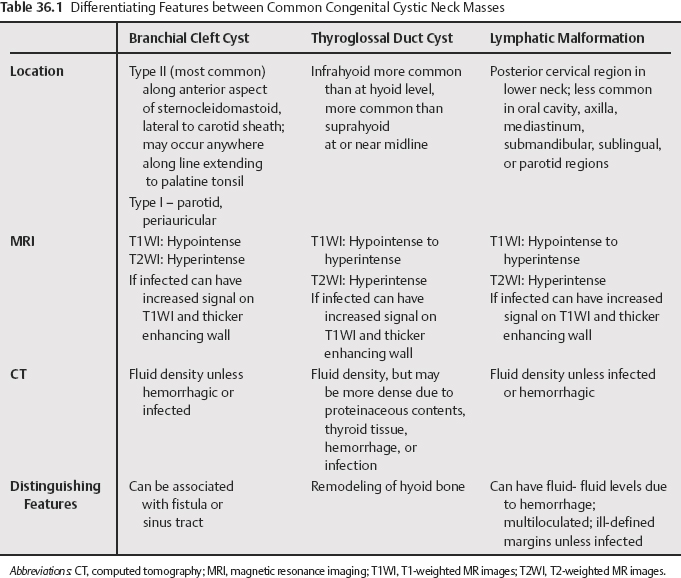
36 The most common congenital cystic lesions of the neck are thyroglossal duct cysts (most common), branchial cleft cysts, and lymphatic malformations (Table 36.1). These lesions arise in remnants of the branchial apparatus, specifically from the cervical sinusof His or cell rests. These lesions typically appear as simple cysts on computed tomography (CT) and magnetic resonance imaging (MRI), with only a thin enhancing wall. The presence of current or prior infection, however, can result in increased density of cyst contents on CT and increased signal intensity on T1-weighted MR images (T1WI), heterogeneity, wall thickening, and irregularity. Branchial cleft anomalies may be cysts, sinuses, or fistulas. They involve the first through fourth branchial clefts, with the second branchial cleft anomalies the most common. First branchial cleft (FBC) anomalies account for 8% to less than 10% of all branchial cleft defects. FBC anomalies occur along a tract extending from the floor of the external auditory canal to the submandibular region. They are classically divided into type I and type II, but probably represent a spectrum of lesions. Type I lesions present as cystic masses. They are generally in a preauricular location, or in or near the parotid gland. Type II lesions present as cysts, sinuses, fistulae, or any combination thereof. They may contain skin adnexal structures and cartilage. Generally, they are located near the angle of the mandible, posterior or inferior to it.
Congenital Cystic Neck Masses
Branchial Cleft Cysts
First Branchial Cleft Cyst
Second Branchial Cleft Cysts
Related posts:

Stay updated, free articles. Join our Telegram channel

Full access? Get Clinical Tree


