Fig. 7.1
Nasolacrimal duct cysts in a 15-day-old girl. (a) Axial T2-weighted image shows cystic masses in the medial canthus of both the orbits. (b, c) Coronal T2-weighted images show the cystic masses extending from the medial canthus into the nasal cavities along the nasolacrimal ducts
7.5.2 Choanal Atresia
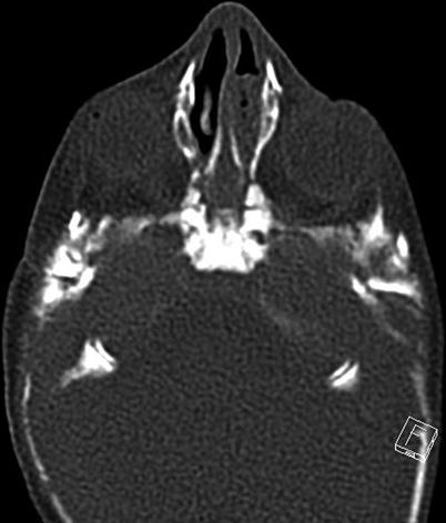
Fig. 7.2
Choanal atresia in a neonate. Axial CT scan with a bone window shows narrowing of both choanae, thickening of the posterior and inferior parts of the vomer, and the presence of an air–fluid level in the left nasal cavity due to choanal obstruction. He also had bilateral external auditory canal atresia
7.5.3 Pyriform Aperture Stenosis
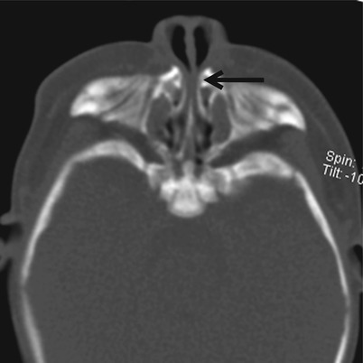
Fig. 7.3
Pyriform aperture stenosis in a 1-month-old infant. Axial CT scan shows inward bowing of the nasal process of the maxilla and narrowing of the anterior nasal cavity (arrow)
7.5.4 Dermoid and Epidermoid Cysts
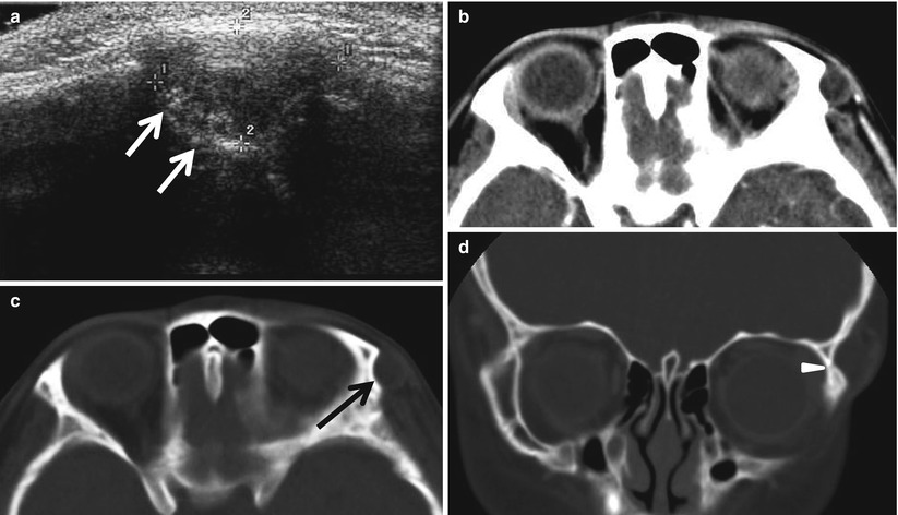
Fig. 7.4
Orbital dermoid cyst in a 5-year-old girl. (a) US shows an oval-shaped nodule with internal echogenic foci in the superolateral wall of the orbit. The underlying bony cortex of the orbital rim produces the appearance of a fossa (arrows). (b) Contrast-enhanced axial CT scan shows a round fatty mass in the superolateral aspect of the left orbit. (c, d) Axial and coronal CT scans of bone window show osseous scalloping that produces the appearance of a fossa (arrow) with a sclerotic margin near the zygomaticofrontal suture (arrowhead)
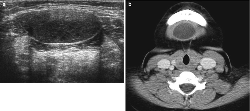
Fig. 7.5
Submental dermoid cyst in a 10-year-old boy. (a) US shows a hypoechogenic cystic mass with internal echogenic lobules and foci. (b) Contrast-enhanced axial CT scan shows a round cystic mass in the midline tongue base. The differential diagnosis includes thyroglossal duct cyst, dermoid cyst, cystic teratoma, and enteric duplication cyst
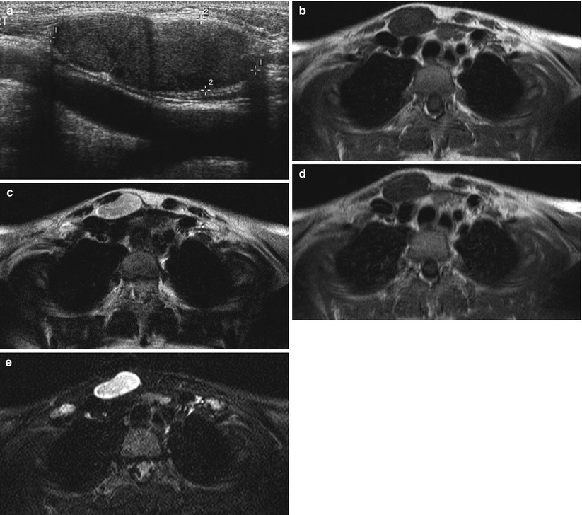
Fig. 7.6
Epidermoid cyst in a 10-year-old girl. (a) US shows a bilobed hypoechoic lesion in the region of the right sternoclavicular junction. Note the internal echogenic foci. (b–d) The mass appears as isosignal intensity on T1-weighted image (b) and slightly high signal intensity on T2-weighted image (c) and demonstrates no contrast enhancement (d). (e) Diffusion-weighted image shows bright high signal intensity of the lesion
7.5.5 Thyroglossal Duct Remnants
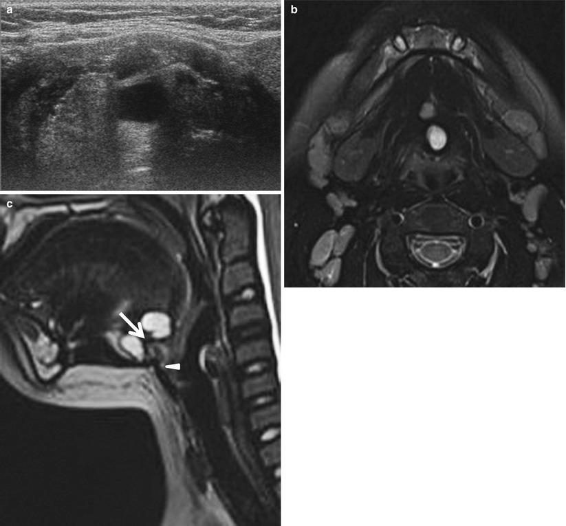
Fig. 7.7
Suprahyoid thyroglossal duct cyst in a 9-year-old boy. (a) US shows a bilocular cystic lesion in the midline aspect of the preepiglottic region. (b) Axial T2-weighted image shows a bilobed cystic mass of high signal intensity in the midline tongue base. (c) Sagittal T2-weighted image shows a fine tract (arrow) between the cystic locules and the relationships between the cyst and the hyoid bone (arrowhead)
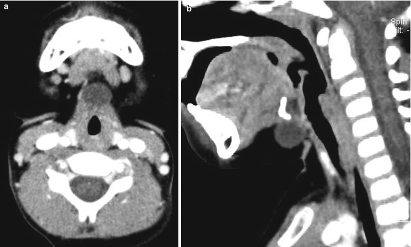
Fig. 7.8
Thyroglossal duct cyst on the level of the hyoid bone in a 3-year-old boy. (a) Contrast-enhanced axial CT scan shows a round midline cyst in the submental space. (b) Sagittal reformatted CT scan shows a well-defined unilocular, thin-walled cyst abutting the hyoid bone
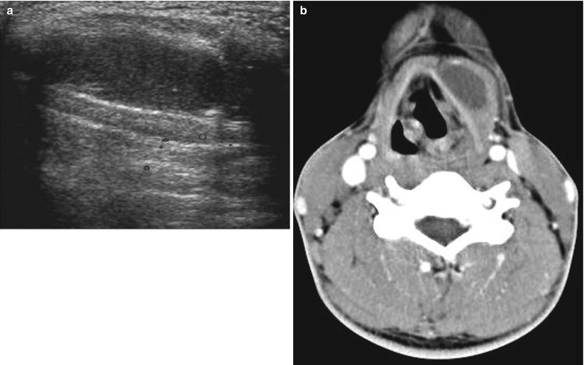
Fig. 7.9
Infrahyoid thyroglossal duct cyst in a 15-year-old boy. (a) US shows a hypoechoic cystic mass embedded in the left strap muscle. (b) Contrast-enhanced axial CT scan shows a well-defined unilocular, thin-walled cyst embedded in the left strap muscle
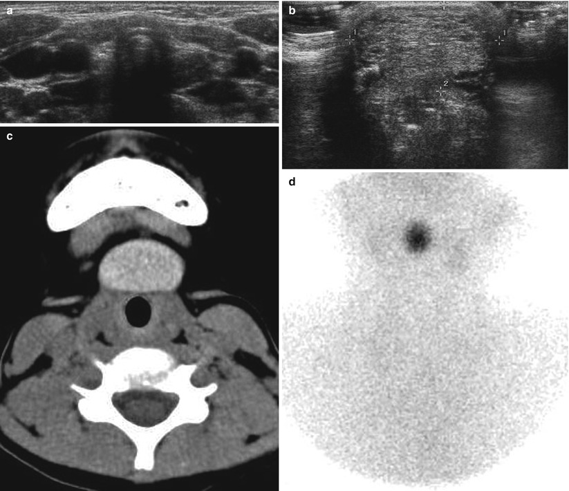
Fig. 7.10
Ectopic thyroid gland in a 7-year-old girl. (a) US shows absence of the thyroid gland at the low neck. (b) US obtained at the level of the hyoid bone shows a heterogeneous echogenic solid mass. (c) Precontrast axial CT scan shows hyperattenuated ectopic thyroid tissue at the level of the hyoid bone. (d) Tc-99m radionuclide scan shows an ectopic lingual thyroid gland and no uptake of the radioisotope at the normal location of the thyroid gland
7.5.6 Branchial Apparatus Anomalies
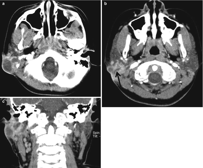
Fig. 7.11
First branchial cleft cyst in an 11-year-old girl. (a, b) Axial CT scans show a complicated, thick-walled cystic mass in the right infra-auricular soft tissue layer with a fine sinus tract (arrow). (c) Coronal reformatted CT scan shows the cystic mass extending just below the ear
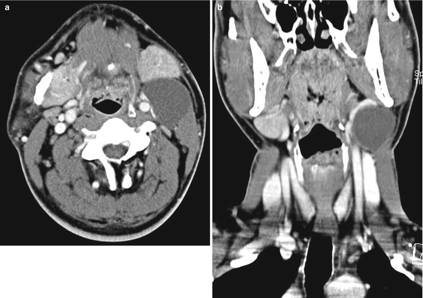
Fig. 7.12
Second branchial cleft cyst in an 18-year-old girl. (a) Axial CT scan shows a thin-walled mass with water attenuation in anteromedial to the sternocleidomastoid muscle. Note the anteriorly displaced submandibular gland, medially displaced left carotid sheath, and posteriorly displaced sternocleidomastoid muscle. (b) Coronal reformatted CT scan shows the typical location of the elongated cyst at the angle of the mandible
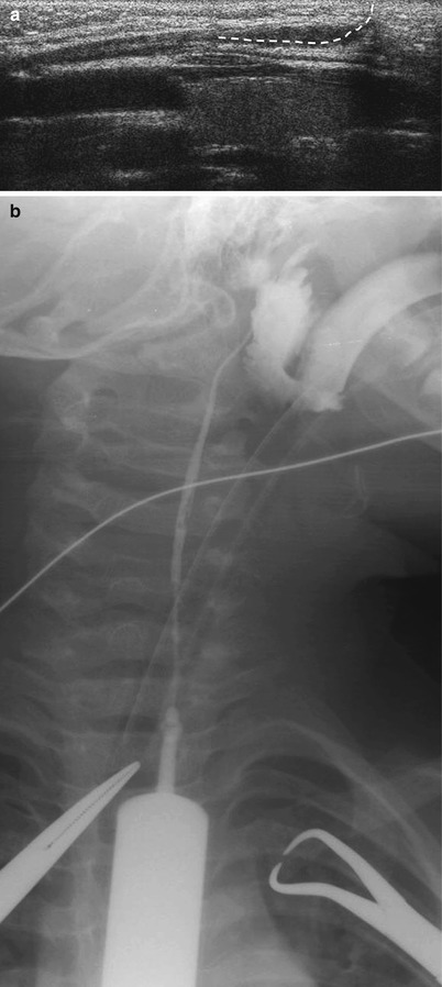
Fig. 7.13
Second branchial cleft fistula in a 2-year-old boy. (a) Longitudinal US of the neck shows a long, linear sinus (—-) from the tiny skin pit on the anterior border of distal sternocleidomastoid muscle. US has a limited role for tracing the entire length of the sinus tract. (b) Operative sinogram shows the complete fistula tract extending from the skin pit in the lower neck to the right tonsillar fossa
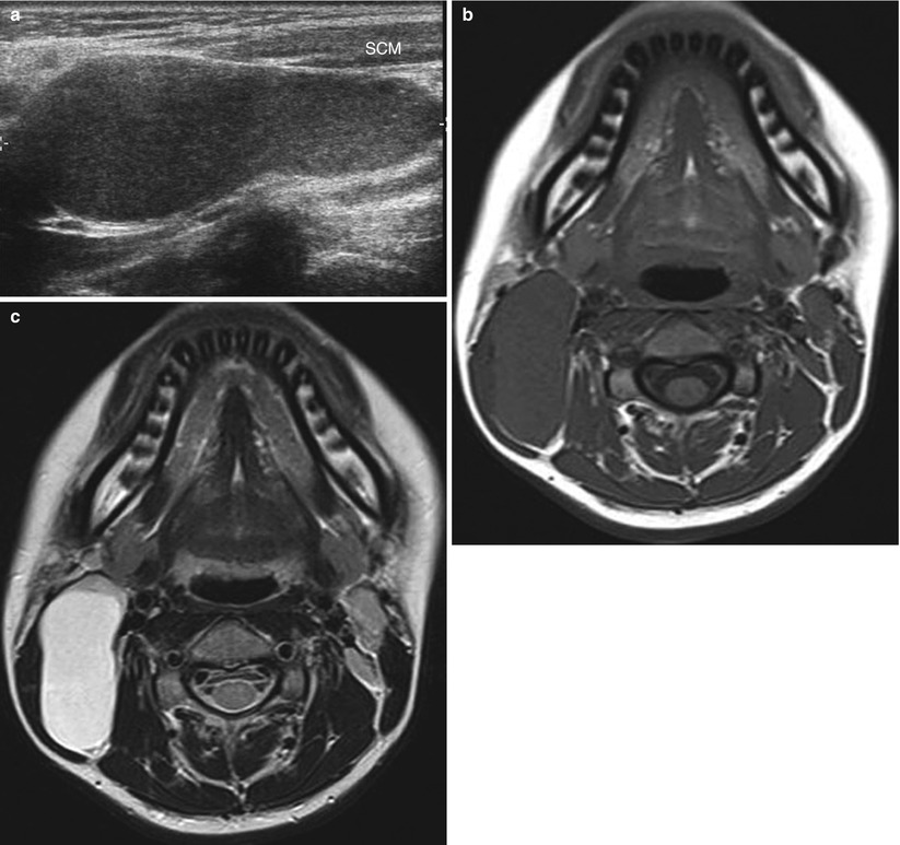
Fig. 7.14


Third branchial cleft cyst in a 16-year-old girl. (a) US shows an elongated cyst along the posterior aspect of the right sternocleidomastoid (SCM) muscle. Note the internal echogenic debridements indicating complicated or proteinaceous content. (b) Axial T1-weighted image shows an elongated cyst of isosignal intensity. (c) Axial T2-weighted image shows the cyst of high signal intensity, posterolateral to the carotid sheath, displacing the sternocleidomastoid muscle anterolaterally
Related posts:


Stay updated, free articles. Join our Telegram channel
Full access? Get Clinical Tree
