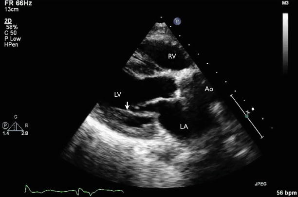
Fig. 9.1
Transthoracic echocardiography (TTE) parasternal long-axis view showing dilatation of the coronary sinus (CS) in the setting of persistent left superior vena cava and mitral regurgitation (arrow). Ao aorta, LA left atrium, LV left ventricle, RV right ventricle
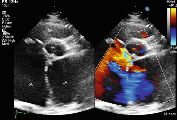
Fig. 9.2
TTE parasternal short-axis view showing atrial septal defect with left-to-right shunt. AV aortic valve, RA right atrium, RV right ventricle
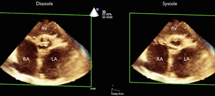
Fig. 9.3
Three-dimensional (3D) TTE short-axis view at the aortic valve level showing the bicuspid aortic valve. The aortic valve orifice appears to have a “fish-mouth” appearance. LA left atrium, RA right atrium, RV right ventricle
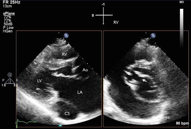
Fig. 9.4
TTE X-plane imaging showing two orthogonal views showing an anterior mitral valve cleft (arrow). Ao aorta, CS coronary sinus, LA left atrium, LV left ventricle, RV right ventricle
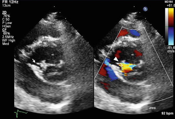
Fig. 9.5
TTE parasternal short-axis view of the mitral valve with color Doppler imaging showing anterior mitral valve cleft (arrow) with corresponding mitral regurgitation (arrowhead)

Fig. 9.6
TTE four-chamber view showing dilated right-sided cardiac chambers with a ventricular upper septal aneurysm. LA left atrium, LV left ventricle, RA right atrium, RV right ventricle
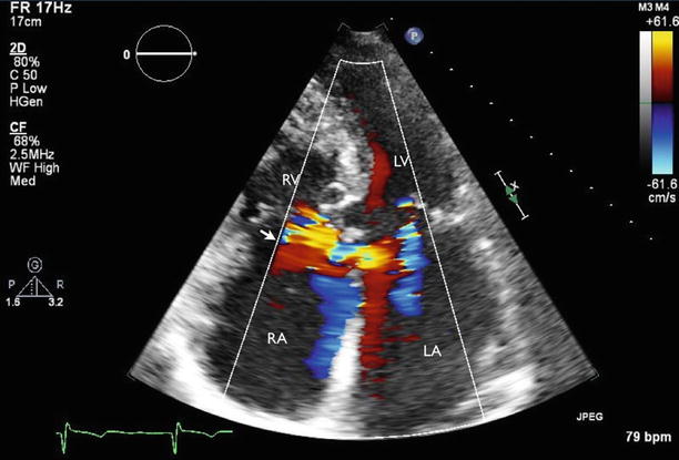
Fig. 9.7
TTE four-chamber view with color Doppler imaging showed atrial septal defect with left-to-right shunt (arrow) at the level close to the cardiac crux, consistent with primum atrial septal defect. Also seen are mitral and tricuspid regurgitation. LA left atrium, LV left ventricle, RA right atrium, RV right ventricle
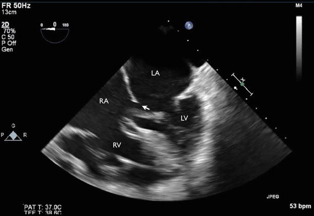
Fig. 9.8
Transesophageal echocardiography (TEE) four-chamber view showed primum atrial septal defect (arrow). LA left atrium, LV left ventricle, RA right atrium, RV right ventricle
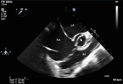
Fig. 9.9
TEE short-axis view at aortic valve level showed bicuspid aortic valve (arrow). LA left atrium, RA right atrium, RV right ventricle
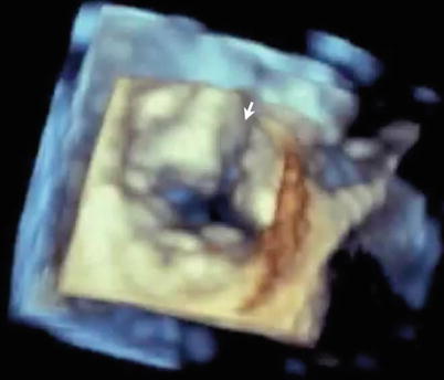
Fig. 9.10
3D TEE of mitral valve (surgical view) showed anterior mitral valve cleft (arrow)
Video 9.1 Transthoracic echocardiography (TTE), parasternal long-axis view, showing dilatation of the coronary sinus in the setting of persistent left superior vena cava and mitral regurgitation (AVI 1923 kb)
Video 9.2 TTE, parasternal short-axis view, showing atrial septal defect with left-to-right shunt (AVI 2013 kb)
Video 9.3 Three-dimensional (3D) TTE short-axis view at the aortic valve level showing the bicuspid aortic valve. The aortic valve orifice appears to have a “fish-mouth” appearance (AVI 1832 kb)
Video 9.4 TTE X-plane imaging showing two orthogonal views showing an anterior mitral valve cleft (AVI 3804 kb)
Video 9.5 TTE parasternal short-axis view of the mitral valve with color Doppler imaging showing anterior mitral valve cleft with corresponding mitral regurgitation (AVI 1818 kb)
Video 9.6 TTE four-chamber view showing dilated right-sided cardiac chambers with a ventricular upper septal aneurysm (AVI 5815 kb)
Video 9.7 TTE four-chamber view with color Doppler imaging showed atrial septal defect with left-to-right shunt at the level close to the cardiac crux, consistent with primum atrial septal defect. Also seen are mitral and tricuspid regurgitation (AVI 2320 kb)
Video 9.8 Transesophageal echocardiography (TEE) four-chamber view showed primum atrial septal defect (AVI 6583 kb)
Video 9.9 TEE short axis view at aortic valve level showed bicuspid aortic valve (AVI 6459 kb)
Video 9.10 3D TEE of mitral valve (surgical view) showed anterior mitral valve cleft (AVI 218 kb)
9.1.1 Learning Points
In ostium primum atrial septal defect, absence of the membranous septum and a cleft in the anterior mitral leaflet are the characteristic features resulting from maldevelopment of the endocardial cushions [1]. The echocardiographic features of mitral valve cleft include the separation of the anterior mitral leaflets during diastole, typically with accessory chordae seen as thin linear echoes between the ridges of both parts of the anterior leaflet and the ventricular septum.
9.2 Case 2. Parachute Mitral Valve
A 27-year-old woman has a history of Shone complex, which includes coarctation of the aorta, a bicuspid aortic valve, and a parachute-variant mitral valve with supramitral ring status post multiple previous operative repairs, including patch arterioplasty, a supramitral ring resection, and subaortic stenosis resection, as well as a Konno procedure with implantation of a #19 St. Jude aortic valve replacement. She is here for routine follow-up. She has remained asymptomatic and continues to exercise vigorously several times per week (Figs. 9.11, 9.12, 9.13, 9.14, and 9.15).