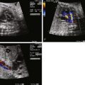
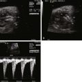
- •
Determine the nature of the heart relationship as being either two separate beating hearts or a single cardiac amalgam.
- •
If two separate hearts, identify the relative size, function, and anatomy of each individual heart.
- •
If a single cardiac amalgam, determine the position and degree of apportionment between the two fetal bodies.
- •
If a single cardiac amalgam identify the number of ventricles, the nature of the communications between the ventricles and the atrioventricular valve connections to the ventricles
- •
If a single cardiac amalgam, identify the venous connections with particular attention to the presence or absence of the inferior vena cava and its position and insertion in the heart.
- •
If a single cardiac amalgam, identify the pulmonic and aortic outflow tracts to each twin and the nature of any obstruction.
Anatomy and Anatomical Associations
Malformations of the heart are common in conjoined twins. Such twins are classified based on the location of connection, with the term -pagus as the suffix, which means “fixed.” Thoracopagus (joined at the thoracic level) are the most common, accounting for 40% of all cases of conjoined twins, followed by omphalopagus (joined at the abdomen predominantly, but may also include the lower thorax) making up 32%. Other forms include pyopagus (joined at the sacrum), ischiopagus (joined at the hip), or craniopagus (joined at the skull). Parapagus is a term used to describe twins fused extensively side to side and can also be known as dicephalus. Cardiac malformations can be seen in any type of twin fusion; however, it is most common in thoracopagus in which there is a 90% incidence of shared pericardium and a 75% incidence of major myocardial connections. The degree of cardiac fusion and the nature of the associated intracardiac malformation determine the potential for surgical separation as well as long-term survival.
The cardiac anatomy of conjoined twins can be quite varied and complex, ranging from two separate hearts and pericardia to hearts that are fused at the atrial or ventricular levels to what is in essence a single cardiac “amalgam” shared by both twins. Coronary arteries and coronary venous drainage may be intertwined and shared. In such fused compound hearts, four is the maximum number of atria and four is the maximum number of ventricles; however, commonly there are fewer of each. One may find a ventricle that is hypoplastic with or without atrioventricular communications leading into it. There can also be multiple communications between the ventricles via ventricular septal defects. The great vessels arising from such a single-heart amalgam to each twin can be abnormally related with conotruncal type anomalies and vessel size discrepancy seen commonly.
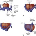
In order to simplify the approach to anatomical evaluation of the heart in conjoined twins, we developed a classification system based on the number of hearts present (type) and the location of the cardiac mass (subclass) ( Figure 34-1 ). This classification system allows for a framework of understanding from which the details of the anatomy can be built upon. In type I, a joined single-heart mass with great vessels arising from opposite poles of the heart perfuse the individual twins. In type II, two distinct heart masses for each twin are present. In subclass A, there is an equal distribution of cardiac mass for each twin; in subclass B, there is an unequal distribution of cardiac mass between the twins. Hence, a thoracopagus with classification IA has a single large heart amalgam compound that is positioned equally between the two twin chests; IIB has a single-heart amalgam compound predominantly positioned within the chest of one twin, but this heart gives rise to vessels that perfuse both twins. In class IIA, each twin has its own separate, relatively equal-sized heart. In addition, each heart may be normal or have an intracardiac abnormality such as a conotruncal anomaly or single-ventricle type congenital heart disease. In class IIB, there are also two separate hearts; however, one is well formed and functions to perfuse the body, whereas the other is primitive and poorly developed. Vascular connections from the twin with the well-formed heart cross over and perfuse its partner.

Although type I with a single-heart amalgam can appear to be a confusing random array of chambers, we have noticed recurring patterns of anatomy. In thoracopagus, the fused twins are typically facing each other. It is best to think of the large heart amalgam in their shared chest as an open book, with the two pages facing you representing the two hearts fused at the center. Often, one can determine a central axis point that distinguishes where the hearts have fused. Typically, there is fusion between the morphologically left ventricles centrally and medially, with the morphologically right ventricles positioned laterally. When there are four ventricles, the two medial left ventricles may share a wall. When there are three or fewer ventricles, the single large central ventricle is typically a fused left ventricle with right ventricles of varying size located on each side. The great vessels can arise from the medial or lateral ventricles but always travel from the ventral conjoined surface of the twins toward the dorsal aspects of each. Pulmonary obstruction or aortic obstruction can be present. Interestingly, when there is vessel obstruction in one twin, the other may have the same type of vessel obstructed or no obstruction. We have not seen any cases in which there was pulmonary obstruction to one twin but aortic obstruction to the other in our fused class I type conjoined heart.
We reviewed a series of 25 conjoined twins evaluated at the Fetal Heart Program at CHOP (unpublished data). Of these, 12 were type IA, 8 type IB, 4 type IIA, and 1 type IIB. Of the type I (single-heart amalgam, n = 20), pulmonary obstruction to both twins was present in 10 (50%), pulmonary obstruction to one twin in 7 (35%), aortic obstruction to both twins in 2 (10%), and no obstruction to either great vessel in 1 twin set (5%). Of the type II (two cardiac masses, n = 5), the hearts were normal in both twins in 2; in 1 set there was transposition of the great vessels in both twins; in 1 set, there was a normal heart in one twin and double-outlet right ventricle in the other; and in 1 set, there was a normal heart in one twin and a rudimentary, primitive single-ventricle heart with complete heart block in the other twin.
Extracardiac abnormalities in conjoined twins are common including congenital diaphragmatic hernia, abdominal wall defects, and imperforate anus. In omphalopagus, a shared liver can be seen, with either one or two bile drainage systems.
Frequency, Genetics, and Development
Conjoined twins occur in the range of 1 in 50,000 to 100,000 pregnancies. Because 60% are stillborn or die shortly after birth, the true incidence is approximately 1 in 200,00 live births. The female to male ratio is 3 : 1.
In the case of conjoined twins, a woman produces only a single egg, which does not fully separate after fertilization. The developing embryo starts to split into identical twins during the first few weeks after conception, but stops before the process is complete. The partially separated egg develops into a conjoined fetus. The twins are, therefore, “identical” with similar genetic makeup. Since in thoracopagus twins, the single heart is multiventricular; it suggests very early union with fusion of the cardiac amalgam before significant differentiation has taken place.
In general, as a consequence of assisted reproductive technologies, the number of twins are increasing, and in combination with improved accuracy of early imaging, conjoined twins are being more readily diagnosed early in pregnancy. Intracytoplasmic sperm injection (ICSI) type of in vitro fertilization has been reported in association with conjoined twinning ; however, the overall numbers of such are too small to declare a cause-and-effect relationship.
Stay updated, free articles. Join our Telegram channel

Full access? Get Clinical Tree


