Crohn Disease
R. Brooke Jeffrey, MD
Key Facts
Terminology
Terminal ileitis, regional enteritis, ileocolitis
Chronic, recurrent, segmental, granulomatous inflammatory bowel disease
Imaging
Best diagnostic clue: Segmental areas of ileo-colonic ulceration and wall thickening on barium study
Early changes seen in barium studies
“Cobblestoning”: Combination of longitudinal and transverse ulcers
Deep fissuring ulcers
Mural thickening: Transmural inflammation, fibrosis
Late changes seen in barium studies
Skip lesions: Segmental/normal intervening areas
“String” sign: Luminal narrowing + ileal stricture
Sinus tracts, fissures, fistulas: Hallmarks of Crohn
Best imaging tools
Barium enema, enteroclysis
MDCT ± contrast
MR for perianal and rectal Crohn disease
Pathology
Possible factors include genetics, environment, infection, psychology; exact etiology unknown
Clinical Issues
Complications: Fistula, sinus, toxic megacolon, obstruction, perforation, malignancy
Diagnostic Checklist
Consider associated findings (cholangitis, arthritis)
CECT: SB wall thickening, mesenteric fat proliferation, hyperemia very suggestive of Crohn
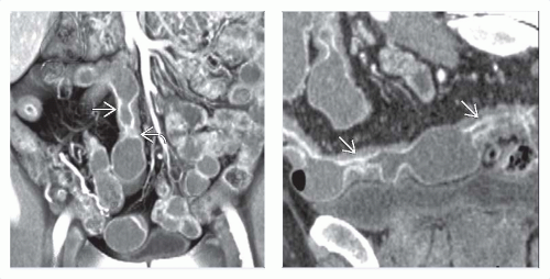 (Left) Coronal volume-rendered CECT enterography in a 27-year-old man presenting with RLQ pain demonstrates marked mucosal hyperemia  and asymmetric mural thickening and asymmetric mural thickening  of the terminal ileum. (Right) Coronal CT enterography curved planar reconstruction through the diseased segment of the terminal ileum in the same patient reveals skip areas of involvement with 2 areas of stricture formation of the terminal ileum. (Right) Coronal CT enterography curved planar reconstruction through the diseased segment of the terminal ileum in the same patient reveals skip areas of involvement with 2 areas of stricture formation  . . |
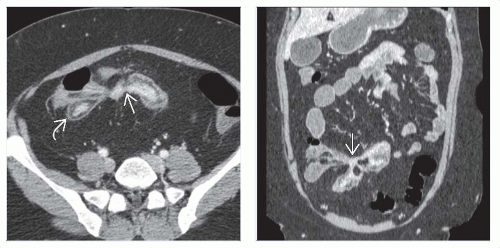 (Left) Axial CECT in a 48-year-old woman presenting with recent weight loss and diarrhea shows segmental small bowel wall thickening characterized by excessive submucosal fat deposition  , indicative of chronic inflammation. The nonepithelialized fistulas , indicative of chronic inflammation. The nonepithelialized fistulas  extend from 1 bowel segment to the others. (Right) Coronal CECT in the same patient again demonstrates the extension of the nonepithelialized fistulas extend from 1 bowel segment to the others. (Right) Coronal CECT in the same patient again demonstrates the extension of the nonepithelialized fistulas  between the bowel segments. between the bowel segments. |
TERMINOLOGY
Synonyms
Terminal ileitis, regional enteritis, ileocolitis
Definitions
Chronic, recurrent, segmental, granulomatous inflammatory bowel disease
IMAGING
General Features
Best diagnostic clue
Segmental areas of ileo-colonic ulceration and wall thickening on barium study
Location
Anywhere along gastrointestinal (GI) tract, from mouth to anus
Most common: Terminal ileum (TI) and proximal colon
Distribution
TI (95%), colon (22-55%)
Rectum (14-50%)
Morphology
Skip lesions (segmental or discontinuous)
Transmural, granulomas (noncaseating type)
“Cobblestone” mucosa, fissures, and fistulas
Fluoroscopic Findings
Barium studies: Early changes
Lymphoid hyperplasia: 1-3 mm mucosal elevations, no ring shadow
“Target” or “bull’s-eye” appearance of aphthoid ulcerations: Punctate shallow central barium collections surrounded by halo of edema
“Cobblestoning”: Combination of longitudinal and transverse ulcers
Deep fissuring ulcers
Mural thickening: Transmural inflammation, fibrosis
Barium studies: Late changes
Skip lesions: Segmental/normal intervening areas
Sacculations seen on antimesenteric border (increased luminal pressure)
Postinflammatory pseudopolyps, haustral loss, intramural abscess
“String” sign: Luminal narrowing and ileal stricture
Sinus tracts, fissures, fistulas are hallmarks of diseaseRelated posts:

Stay updated, free articles. Join our Telegram channel

Full access? Get Clinical Tree


