5
RADIATION DAMAGE REPAIR AND MODELS FOR NORMAL TISSUES
ADITYA JULOORI, CHOAMEI XIANG, MICHAEL A. WELLER, AND JENNIFER S. YU
Question 1
What is the difference between lethal damage, potentially lethal damage, and sublethal damage?
Question 3
What is the importance of the shoulder width on traditional curves for normal tissues?
Question 1 What is the difference between lethal damage, potentially lethal damage, and sublethal damage?
Answer 1
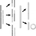
Lethal damage from radiation is not reversible or reparable and leads to cell death. Potentially lethal damage can be modified by the conditions cells are placed in after being irradiated (observed in cell culture conditions). For example, this has been demonstrated in vitro by putting cells in saline for 6 hours after irradiation in order to plateau the growth curve—these cells then had a higher surviving fraction after radiotherapy than those cells that were not placed in these conditions after radiotherapy. It is hypothesized that tumors with relatively radioresistant histology like melanoma are able to repair this potentially lethal damage. Sublethal damage can be repaired in hours under normal conditions.
Hall EJ, Giaccia AJ. Radiation carcinogenesis. In: Hall EJ, Giaccia AJ, eds. Radiobiology for the Radiologist. 7th ed. Philadelphia, PA: Lippincott Williams & Wilkins; 2006:135–153.
Question 3 What is the importance of the shoulder width on traditional curves for normal tissues?
Answer 3
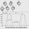
Various normal tissues have a wide range of radiosensitivities, with the shoulder length on survival curves being the principal variable. For example, jejunal crypt cells have a large shoulder while bone marrow stem cells have a limited shoulder. The shoulder length is larger for some tissues because of the inherent ability to repair potentially lethal damage.
Hall EJ, Giaccia AJ. Radiation carcinogenesis. In: Hall EJ, Giaccia AJ, eds. Radiobiology for the Radiologist. 7th ed. Philadelphia, PA: Lippincott Williams & Wilkins; 2006:135–153.
Question 5
What is Casarett’s classification of tissue radiosensitivity based on?
Question 7
What is an F-type population according to the Michalowski classification?
Question 5 What is Casarett’s classification of tissue radiosensitivity based on?
Answer 5
Cells are divided into four major categories based on histopathological observations, from most sensitive to least resistant. Group I cells divide regularly with no differentiation (e.g., intestinal crypt cells). Group II cells divide regularly with some differentiation between divisions (e.g., myelocytes). Group III cells do not divide regularly and are variably differentiated (e.g., liver). Group IV cells do not divide and are highly differentiated (e.g., nerve cells).
Hall EJ, Giaccia AJ. Radiation carcinogenesis. In: Hall EJ, Giaccia AJ, eds. Radiobiology for the Radiologist. 7th ed. Philadelphia, PA: Lippincott Williams & Wilkins; 2006:135–153.
Rubin P, Casarett GW. Clinical Radiation Pathology. Vol. 1. Philadelphia, PA: WB Saunders; 1968.
Question 7 What is an F-type population according to the Michalowski classification?
Answer 7
These tissues follow a “flexible” model. These tissues are in organs where the cells do not typically divide unless triggered to do so in response to damage. Examples of this type of tissue include liver, skin, and thyroid. These tissues have no hierarchy and no compartments.
Wheldon TE, Michalowski AS. Alternative models for the proliferative structure of normal tissues and their response to irradiation. Br J Cancer. 1986;7(suppl):382–385.
Question 9
How has the dose–response relationship for radiation exposure to skin been studied?
Related posts:
 of CancerKailin Yang and Jennifer S. Yu
Influencing Tumor Control and ComplicationsDaesung Lee
Radiation Therapy ModalitiesSusan Kost and Andrew Godley
of CancerKailin Yang and Jennifer S. Yu
Influencing Tumor Control and ComplicationsDaesung Lee
Radiation Therapy ModalitiesSusan Kost and Andrew Godley
 Chromosome and Chromatid Damage, Repair, and MeasurementEhsan H. Balagamwala, C. Marc Leyrer, Jeffrey A. Kittel, and Alex Almasan
 and Risks in Nonradiation Oncology SubspecialtiesFrank Dong, Kevin Wunderle, and Salim Balik
 KineticsMatthew C. Ward and Richard Blake Ross
Stay updated, free articles. Join our Telegram channel

Full access? Get Clinical Tree


