Fig. 4.1
Transesophageal 3D reconstruction of the mitral valve demonstrating prolapse of the middle and lateral posterior leaflet scallops (P1 & P2)
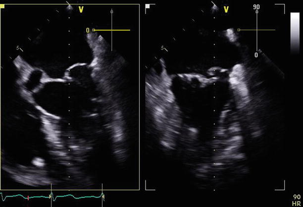
Fig. 4.2
Transesophageal echo biplane (0 and 90°) view of the mitral valve demonstrating prolapse of P2 & P2
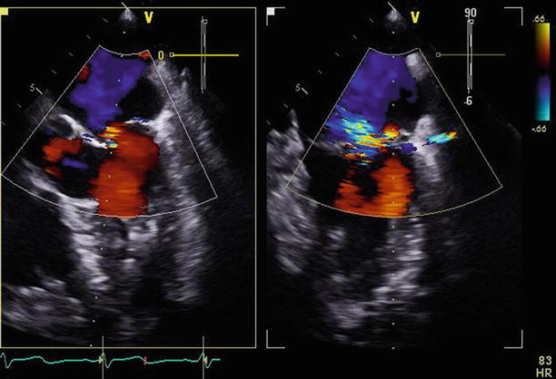
Fig. 4.3
The same views with color Doppler, demonstrating eccentric, anteriorly directed mitral regurgitation (MR), which was classified as 3–4+. Regurgitation can be difficult to quantitate on one view because of the eccentricity of the jet
Video 4.1 Transesophageal 3D reconstruction of the mitral valve demonstrating prolapse of the middle and lateral posterior leaflet scallops (P1 & P2) (AVI 950 kb)
Video 4.2 Transesophageal echo biplane (0 and 90°) view of the mitral valve demonstrating prolapse of P2 & P2 (AVI 5627 kb)
Video 4.3 The same views with color Doppler, demonstrating eccentric, anteriorly directed mitral regurgitation, which was classified as 3–4+ (AVI 1477 kb)
4.2 Case 2. Mitral Valve Posterior Leaflet Flail
A 74-year-old man presented with increasing fatigue on a background history of inferior wall myocardial infarction, which had been medically managed 15 years earlier. Echocardiogram revealed a flail posterior mitral valve leaflet with severe mitral regurgitation and moderately elevated pulmonary pressures. He was referred for a mitral valve repair and a bypass graft to the right coronary artery (Figs. 4.4, 4.5, 4.6, 4.7, 4.8, and 4.9).
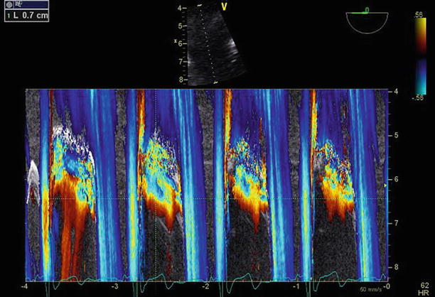
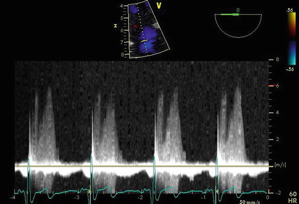
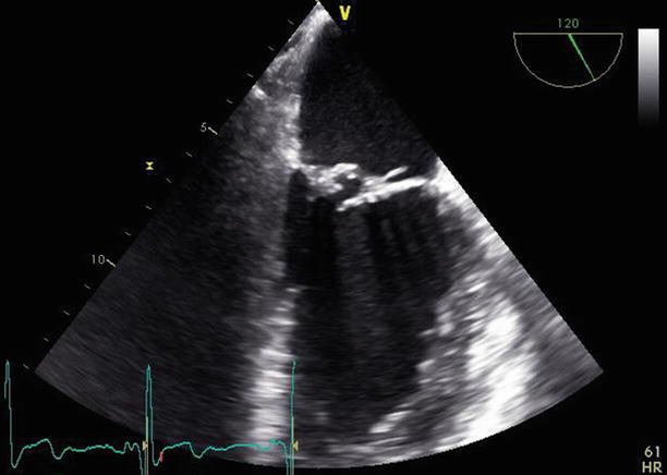
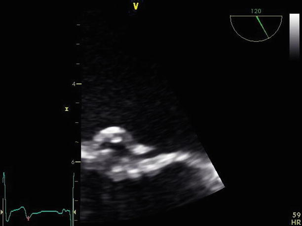
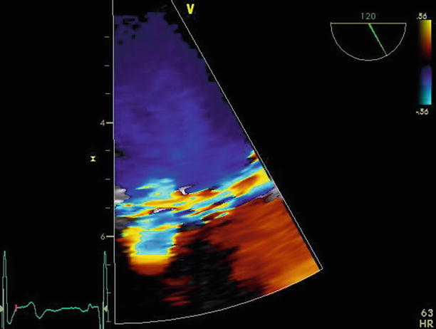
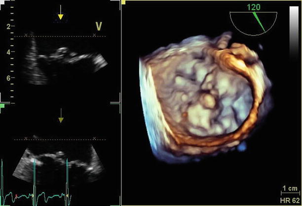
Fig. 4.4
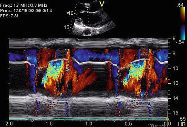
Color M-mode through the mitral valve, demonstrating predominantly early-systolic and late-systolic MR
Fig. 4.5
Continuous-wave Doppler through the mitral valve, demonstrating predominantly early-systolic and late-systolic MR
Fig. 4.6
Transesophageal 120° view confirms flail involving the middle posterior leaflet scallop (P2)
Fig. 4.7
Transesophageal 120° view, zooming up on the flail middle posterior leaflet scallop (P2)
Fig. 4.8
Color Doppler (transesophageal 120° view) demonstrating severe, anteriorly directed MR. Note wide vena contracta; proximal isovelocity surface area (PISA) measurements likely underestimate severity of MR owing to jet eccentricity
Fig. 4.9
Three-dimensional (3D) reconstruction of the mitral valve demonstrating the flail middle posterior leaflet scallop (P2)
Video 4.4 Transesophageal 120° view confirms flail involving the middle posterior leaflet scallop (P2) (AVI 7312 kb)
Video 4.5 Transesophageal 120° view, zooming up on the flail middle posterior leaflet scallop (P2) (AVI 6013 kb)
Video 4.6 Color Doppler (transesophageal 120° view) demonstrating severe, anteriorly directed MR. Note wide vena contracta; proximal isovelocity surface area (PISA) measurements likely underestimate severity of MR owing to jet eccentricity (AVI 1247 kb)
Video 4.7 Three-dimensional (3D) reconstruction of the mitral valve demonstrating the flail middle posterior leaflet scallop (P2) (AVI 1144 kb)
4.3 Case 3. Posterior Mitral Valve Leaflet Prolapse After Previous Mitral Valve Repair
A 46-year-old male physician presented for surgical evaluation of mitral regurgitation (Carpentier type 2, severe mitral regurgitation secondary to posterior leaflet prolapse), on a background history of previous mitral valve repair several years earlier. He had remained essentially asymptomatic, but serial monitoring via echocardiography revealed an increasing effective regurgitant orifice area of 0.94 cm2, regurgitant volume of 124 mL, severely dilated left and increasing pulmonary pressures (estimated right ventricular systolic pressure of 51 mmHg) (Figs. 4.10, 4.11, 4.12, and 4.13).
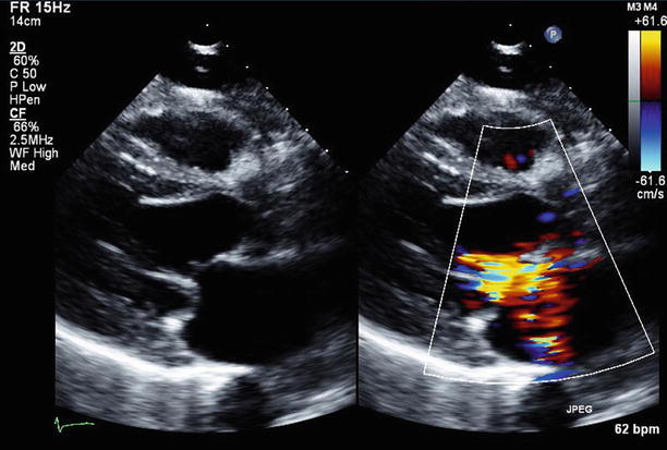
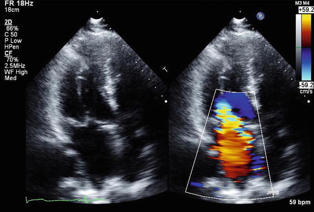
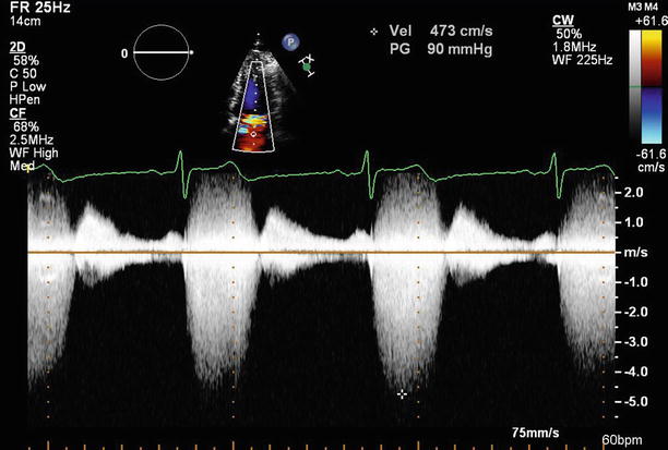
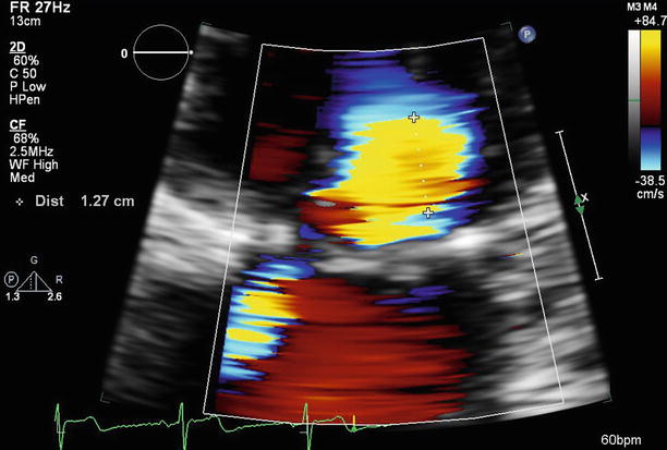
Fig. 4.10
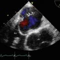
Parasternal long-axis view in two-dimensional (2D) and color Doppler imaging demonstrating posterior leaflet prolapse with eccentric, anteriorly directed mitral regurgitation
Fig. 4.11
Apical long-axis view demonstrating that the anterior mitral valve regurgitation is severe
Fig. 4.12
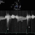
Continuous wave Doppler demonstrates the mitral regurgitant jet. Note the jet density and holosystolic profile, which supports the conclusion that the regurgitation is severe. The peak jet velocity may be underestimated because of the jet’s eccentric nature; it is difficult to ensure that the transducer beam is parallel to the regurgitant jet
Fig. 4.13
From a PISA diameter of 1.27 cm, a mitral valve Vmax of 473 cm/s, and an aliasing velocity of 61.6 cm/s (see Fig. 4.12), the estimated regurgitant orifice area is 1.3 cm2, which is classified as severe
Video 4.8 Parasternal long-axis view in two-dimensional (2D) and color Doppler imaging demonstrating posterior leaflet prolapse with eccentric, anteriorly directed mitral regurgitation (AVI 2643 kb)
Video 4.9 Apical long-axis view demonstrating that the anterior mitral valve regurgitation is severe (AVI 3332 kb)
4.4 Case 4. Bileaflet Mitral Valve Prolapse and Flail
A 62-year-old man with known myxomatous mitral valve degeneration and secondary mitral insufficiency presented for consideration of minimally invasive mitral valve surgery. He had a longstanding history of heart murmur. Echocardiographic findings included severe mitral insufficiency, with an estimated regurgitant orifice area of 1.28 cm2, mild pulmonary hypertension, and left ventricular dilatation (end-diastolic dimension of 5.9 cm), but his left ventricular systolic function was preserved, with an ejection fraction of 64 %. Although he was asymptomatic, his echocardiographic findings supported a recommendation for mitral valve surgery because of the regurgitation severity and the likelihood of a successful repair (AHA/ACC Class IIa) (Figs. 4.14, 4.15, 4.16, 4.17, 4.18, 4.19, 4.20, and 4.21).
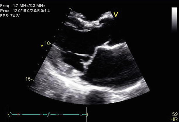
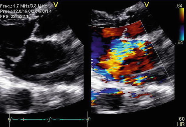
Fig. 4.14
Parasternal long-axis imaging of the mitral valve demonstrates bileaflet mitral valve prolapse
Fig. 4.15
Comparison with color Doppler imaging reveals severe central and anteriorly directed mitral regurgitation