CHAPTER 18 Dehiscent Jugular Bulb
Epidemiology
In the literature, the reported incidence of a dehiscent jugular bulb varies from 2 to 7%.
Clinical Features
Dehiscence of the jugular bulb may be seen as a bluish vascular retrotympanic mass in the posteroinferior quadrant of the tympanic membrane, when viewed with an otoscope. This may present with venous pulsatile tinnitus or as an asymptomatic mass that cannot be differentiated from a paraganglioma clinically. Associated conductive hearing loss secondary to ossicular impingement is rare.
Pathology
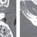
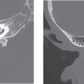
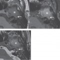
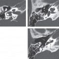
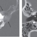
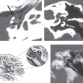
A dehiscent jugular bulb is a congenital variant that can present as a vascular pseudomass in the middle ear. Dehiscence is defined by absence of the bony canal wall between the lateral aspect of the jugular foramen and middle ear cavity.
The jugular bulb variations that commonly occur include an asymmetrically large jugular bulb, a high-riding jugular bulb with or without dehiscence, and a jugular diverticulum. An asymmetrically large jugular bulb is a common finding and is not thought to be a cause of symptoms. In the asymmetrically large jugular bulb, the right jugular bulb is larger twice as often as on the left side.
Stay updated, free articles. Join our Telegram channel

Full access? Get Clinical Tree


