(1)
Kaiser Permanente, Southern California Permanente Medical Group, Riverside, CA, USA
(2)
Molecular Imaging Center and PET Clinic, University of Southern California, Los Angeles, CA, USA
(3)
Keck School of Medicine, University of Southern California, Los Angeles, CA, USA
Case 13.1: Alzheimer’s Dementia
History
67-year-old female with cognitive impairment.
Findings
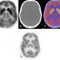
There is bilaterally decreased temporal and posterior parietal cortical activity such that it is less than the cerebellar cortical activity (Fig. 13.1). There is sparing of the motor strips, basal ganglia, and visual. Age-appropriate cortical atrophy is noted. There is no evidence of ischemia or mass lesion.


Fig. 13.1
Impression
Abnormal metabolism consistent with dementia of Alzheimer’s type.
Pearls and Pitfalls
Alzheimer’s disease (AD) is the most common form of dementia. Association cortices are more severely involved, while the primary somatosensory and motor cortices, the basal ganglia, the thalamus, and the cerebellum are relatively spared.
FDG-PET demonstration of the classic metabolic abnormality associated with pathologically verified AD has a sensitivity of 93 %, a specificity of 63 %, and an accuracy of 82 %.
Discussion
It was first described by German psychiatrist and neuropathologist Alois Alzheimer in 1906 and was named after him. Many causes of dementia symptoms exist. Alzheimer’s disease is the most common cause of a progressive dementia characterized with gradual decline in cognition and behavior. Accurate early diagnosis of AD is important because early use of medications may improve or delay the cognitive loss that occurs in mild-to-moderate disease. Normal aging of the brain is characterized by a regional decline in the cerebral glucometabolism of the prefrontal cortex (Fig. 13.1).
Stay updated, free articles. Join our Telegram channel

Full access? Get Clinical Tree


