Presentation and Presenting Images
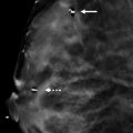
( ▶ Fig. 26.1, ▶ Fig. 26.2, ▶ Fig. 26.3, ▶ Fig. 26.4)
A 59-year-old female with a history of right breast cancer treated 5 years ago presents for screening mammography.
26.2 Key images
26.2.1 Breast Tissue Density
There are scattered areas of fibroglandular density.
26.2.2 Imaging Findings
In the upper outer quadrant of the left breast at the 1 o’clock location in the posterior depth, there is a focal asymmetry. This is best seen on the digital breast tomosynthesis (DBT) images and especially the mediolateral (MLO) DBT images.
26.3 BI-RADS Classification and Action
Category 0: Mammography: Incomplete. Need additional imaging evaluation and/or prior mammograms for comparison.
26.4 Diagnostic Images
( ▶ Fig. 26.7, ▶ Fig. 26.8, ▶ Fig. 26.9, ▶ Fig. 26.10, ▶ Fig. 26.11, ▶ Fig. 26.12)
26.4.1 Imaging Findings
The diagnostic imaging demonstrates a persistent irregular mass with indistinct margins at the 1 o’clock location in the posterior depth. Targeted ultrasound reveals a 4 × 3 × 4 mm irregular mass with indistinct margins and posterior acoustic shadowing. This mass was assessed to be suspicious and a biopsy recommended. The postbiospy craniocaudal (CC) and mediolateral (ML) mammogram images ( ▶ Fig. 26.11 and ▶ Fig. 26.12) demonstrate the biopsy clip at the site of the mass seen mammographically.
26.5 BI-RADS Classification and Action
Category 4B: Moderate suspicion for malignancy
26.6 Differential Diagnosis
Fibromatosis: This mass is irregular with an ultrasound correlate. There is considerable overlap of characteristic imaging findings between fibromatosis and breast cancer, therefore a biopsy is necessary. A biopsy yielding fibromatosis would be concordant with the mammographic, DBT, and ultrasound findings.
Carcinoma: This mass is irregular in appearance and would require a biopsy to exclude breast cancer as the diagnosis.
Normal breast tissue as a summation artifact: This small focal asymmetry persists on additional imaging and is new compared to prior exams (not shown). Thus, this finding should not be dismissed but rather evaluated.
26.7 Essential Facts
Fibromatosis is a stromal tumor.
Fibromatosis is rare, accounting for less than 0.2% of all breast tumors.
Fibromatosis is benign, but locally aggressive. These tumors require wide local excision to prevent recurrence.
Fibromatosis tumors typically present mammographically as spiculated masses.
Fibromatosis significance in breast imaging is that it can mimic breast cancer.
These tumors are more often located near the chest wall.
Magnetic resonance (MR) imaging may be helpful to evaluate the extent of the tumor and the possibility of chest-wall invasion if the tumor is not fully seen on the mammogram.
26.8 Management and Digital Breast Tomsynthesis Principles
Technically DBT reduction of overlapping tissue effects make masses with indistinct or spiculated margins more conspicuous. Thus, fibromatosis, along with cancers, will be more readily apparent on DBT.
The imaging features of a spiculated locally invasive tumor make it suspicious for malignancy and not distinguishable from breast cancer without a biopsy.
26.9 Further Reading
[1] Glazebrook KN, Reynolds CA. Mammary fibromatosis. AJR Am J Roentgenol. 2009; 193(3): 856‐860 PubMed

Fig. 26.1 Left craniocaudal (LCC) mammogram.
Stay updated, free articles. Join our Telegram channel

Full access? Get Clinical Tree


