and Zdeněk Fryšák1
(1)
Department of Internal Medicine III – Nephrology, Rheumatology and Endocrinology, Faculty of Medicine and Dentistry, Palacky University Olomouc and University Hospital Olomouc, Olomouc, Czech Republic
Keywords
GoiterPalpationUltrasoundThyroid volume2.1 Essential Facts
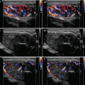
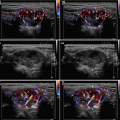
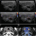
Diffuse goiter is a generalized thyroid hyperplasia. When measured thyroid volume (Tvol) by US, the goiter is defined by Gutekunst from the year 1988 as Tvol >18 ml in women and Tvol >25 ml in men (Figs. 2.1 and 2.2) [1].
Normative data of Tvol, showing variations with age, regional factors, and iodine status of the population, have been reported in different populations.
Iodine deficiency endemic area is defined by the goiter prevalence and the median urinary iodine concentration in a population. The local sex-specific reference values at different ages and body surface areas are not a constant proportion of the WHO/ICCIDD-recommended reference. A further limitation of the WHO/ICCIDD-recommended reference is the lack of normative values for children with small body surface areas (<0.8 m2) commonly found in developing countries [2].
Epidemiologic study for thyroid abnormalities were performed by Rallison et al. in 4819 school-age children (SAC), ages 11–18 years, from 1965 to 1968. Two-thirds of this original cohort (3121) were re-examined 20 years later, in 1985 and 1986.
In the initial examinations (1965–1968), 185 thyroid abnormalities were found (3.7% of the total). These abnormalities were, in order from the most common to the least common: diffuse hypertrophy with normal function (adolescent goiter) in 19.3/1000; CLT in 12.7/1000; and thyroid nodules in 4.6/1000, including two papillary thyroid carcinomas (PTC). Hyperthyroidism or hypothyroidism was found in 1.9/1000.
In the follow-up examinations (1985–1986), 298 (10.5%) subjects had thyroid abnormalities (10.5% of the total). These abnormalities were, in order from the most common to least common: CLT in 51.3/1000; simple goiters in 28.7/1000; and nodules in 23.2/1000, which included 10 carcinomas. In addition, 9/1000 had hypothyroidism and 3.9/1000 had hyperthyroidism [3].
Of the 92 subjects with simple or adolescent goiter in 1965–1968, 60% were normal by 1985–1986, 20% were unchanged, approximately 10% had developed CLT, and approximately 3% had developed colloid goiters. Of 61 subjects with CLT, 27% had become normal, about 33% remained unchanged, and 33% had become hypothyroid [3].
In a study by Hintze et al., a palpation (WHO 1960 palpation criteria) and an ultrasound investigation of the thyroid were performed on 569 unselected elderly subjects greater than 60 years of age from the general population of an iodine-deficient area. Of the subjects, 489 were female and 80 were male.
By palpation, no thyroid enlargement was noticed in 54% of the subjects; goiter IA (palpable lobes larger than the terminal phalanges of the subject’s thumbs) in 18%; goiter IB (visible with the neck extended) in 17%; goiter II (visible with the head in normal position) in 9%; and goiter III (visible at a distance) in 2%.
Tvol (median) by US was 18.6 ml for the entire group (19.2 ml median in the women and 16.6 ml median in the men). 18% had thyroid nodules and 8% had cystic lesions. The goiter prevalence of 54% in women (>18 ml) and of 23% in men (>25 ml) was obtained. Subjects with goiter demonstrated a significantly lower thyrotropin (TSH) value and a higher thyroglobulin value.
In summary, the study shows a high prevalence of goiter in elderly subjects, a high prevalence of nodules in these thyroids, a negative correlation of goiter volume with TSH, and a positive correlation of goiter volume with the thyroglobulin concentration [4].
Hormonal changes and metabolic demands during pregnancy result in profound alterations in the biochemical parameters of thyroid function:
a marked increase in serum thyroxine-binding globulin levels.
a marginal decrease in free hormone concentrations (in iodine-sufficient conditions) that is significantly amplified if there is iodine restriction or overt iodine deficiency.
a frequent trend toward a slight increase in basal TSH values between the first trimester and the delivery.
a direct stimulation of the maternal thyroid gland by elevated levels of human chorionic gonadotropin (hCG), which occurs mainly near the end of the first trimester and can be associated with a transient lowering of serum TSH. Goiters formed during gestation may only partially regress after parturition.
Pregnancy, therefore, represents one of the environmental factors that may explain the higher prevalence of goiter and thyroid disorders in the female population. An iodine-deficient status in the mother also leads to goiter formation in the progeny. When adequate iodine supplementation is given early during pregnancy, it allows for the correction and almost complete prevention of maternal and neonatal goiterogenesis. The ideal dietary allowance of iodine recommended by the WHO is 200 μg/day for pregnant women [5]. The iodine requirement increases during pregnancy and current recommended intake is in the range of 220–250 μg/day [6].
In 1997 the WHO/ICCIDD proposed new references for Tvol in SAC that were based on data from children in iodine-replete countries (Netherlands, Slovakia, France, and Austria). Table 2.1 presents the upper normal limit for Tvol, by age. However, in countries with a high prevalence of child growth retardation, the limits for Tvol shown are unsuitable. In such cases, Tvol is provisionally considered to be more directly a function of total body surface area (Table 2.2) [7].
However, subsequent reports suggested that the 1997 WHO/ICCIDD references were overestimating the real values. Data of Tvol (medians and P97s) reported in 1999–2000 in iodine-sufficient SAC (United States, Switzerland, and Malaysia) were distinctly lower than those in the European children from the 1997 data. The larger Tvol in the 1997 reference data may have been a residual effect of the iodine deficiency that existed in many European countries up to the early 1990s. Moreover, in 2000 a WHO/ICCIDD workshop on thyroid ultrasound uncovered a large systematic measurement bias in the 1997 references; +30% Tvol at all ages and all body surface areas [8].
The latest study by Zimmermann, published in 2004, used validated techniques to measure Tvol by US. The subjects were 3500 healthy SAC (6–12 years old) living in six sites on five continents of longstanding iodine sufficiency (Switzerland, Japan, United States, South Africa, Peru, and Bahrain). Compared with previous WHO data for Tvol in SAC from 1997, these new data are more conservative (Tables 2.3 and 2.4). For example, for the boys, both the age- and BSA-specific P97 values for Tvol in this sample is 20% smaller than the corrected 1997 reference values and 15 to 20% smaller than the 1993 WHO reference values. The 1993 (Swedish and German children) and 1997 WHO reference values were based on data from European children. These new international reference values for Tvol by US can be used for goiter screening in the context of IDD monitoring [9].
Table 2.1
Upper limit of normal thyroid volume (Tvol) measured by US in iodine-replete children aged 6–15 years, as a function of age, according WHO 1997 [7]
Age (years) | Boys Tvol (ml) | Girls Tvol (ml) |
|---|---|---|
6 | 5.4 | 5.0 |
7
Related posts:Stay updated, free articles. Join our Telegram channel
Full access? Get Clinical Tree
 Get Clinical Tree app for offline access

|