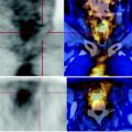
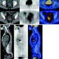
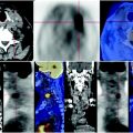
Fig. 59.1
In correspondence of the body and tail of the pancreas CT-PET shows a coarse expansive, diffusely inhomogeneous solid mass, which infiltrates locoregional tissues, in particular lymph nodes, especially celiac-mesenteric and retroperitoneal ones. This mass has a high FDG metabolism. There are multiple peritoneal nodules characterized by abnormal glucose consumption. Conspicuous neoplastic ascites. Obstructive expansion of intra- and extra-hepatic biliary ducts
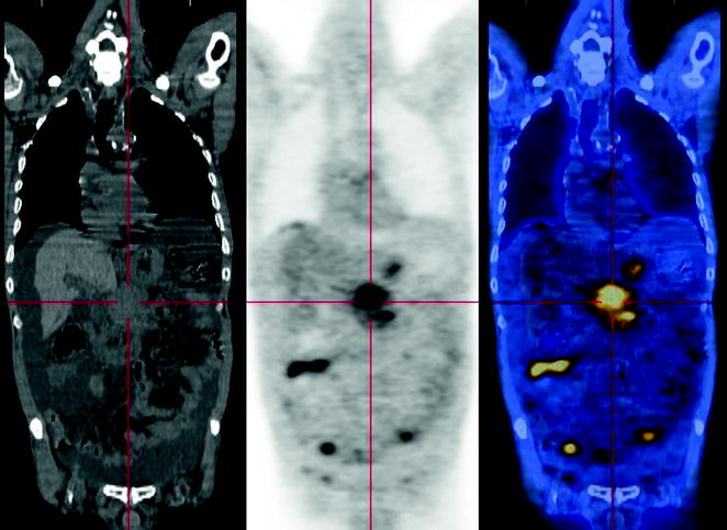
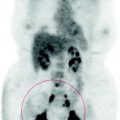
Fig. 59.2


The PET scan shows a tumor of the pancreas with a high metabolism with nodal metastases, peritoneal carcinomatosis and secondary ascites. Moderate obstructive dilatation of the biliary tract
Stay updated, free articles. Join our Telegram channel

Full access? Get Clinical Tree


