Disorders of the Elbow
Anterior
Introduction
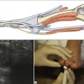
It is important to identify both the biceps and brachialis tendons in the antecubital fossa as, if only a single tendon is present, it means that the other, usually the biceps, is ruptured (Fig. 8.1).
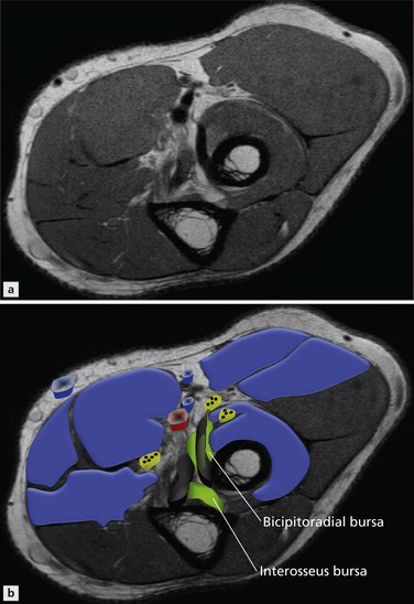
Apart from rupture, the principal pathological processes that affect biceps are tendinopathy, cubital and interosseous bursitis. Tendinopathy of brachialis is rare.
Biceps Tendinopathy
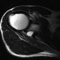
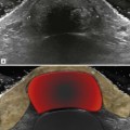
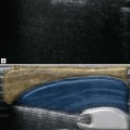
In the earliest stages, fluid gathers within the tendon sheath and synovial thickening occurs. The tendon remains normal initially with a typical reticular reflective pattern best appreciated on axial images, but also when good long-axis images are obtained (Fig. 8.2). Ultimately internal signal changes representing focal tendon degeneration lead to delamination and partial tears (Fig. 8.3). A hypertrophic pattern is more usual and the combination of tendon hypertrophy, fibre disorganization, fluid distension of the tendon sheath and increased Doppler signal is typical.
Stay updated, free articles. Join our Telegram channel

Full access? Get Clinical Tree


